1MS6 | pdb_00001ms6

Dipeptide Nitrile Inhibitor Bound to Cathepsin S.


Experimental Data Snapshot

  • Method: X-RAY DIFFRACTION
  • Resolution: 1.90 Å
  • R-Value Free: 
    0.197 (Depositor) 
  • R-Value Work: 
    0.155 (Depositor) 
  • R-Value Observed: 
    0.162 (Depositor) 

wwPDB Validation 3D Report Full Report

Validation slider image for 1MS6

This is version 1.3 of the entry. See complete history

Literature

Design and synthesis of dipeptide nitriles as reversible and potent Cathepsin S inhibitors

Ward, Y.D.Thomson, D.S.Frye, L.L.Cywin, C.L.Morwick, T.Emmanuel, M.J.Zindell, R.McNeil, D.Bekkali, Y.Giradot, M.Hrapchak, M.DeTuri, M.Crane, K.White, D.Pav, S.Wang, Y.Hao, M.H.Grygon, C.A.Labadia, M.E.Freeman, D.M.Davidson, W.Hopkins, J.L.Brown, M.L.Spero, D.M.

(2002) J Med Chem 45: 5471-5482

  • DOI: https://doi.org/10.1021/jm020209i
  • Primary Citation Related Structures: 
    1MS6

  • PubMed Abstract: 

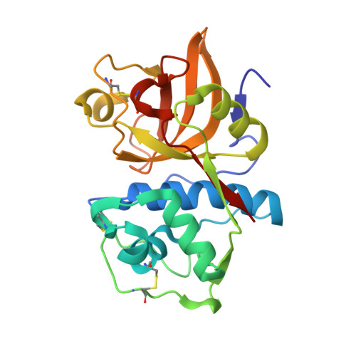
    The specificity of the immune response relies on processing of foreign proteins and presentation of antigenic peptides at the cell surface. Inhibition of antigen presentation, and the subsequent activation of T-cells, should, in theory, modulate the immune response. The cysteine protease Cathepsin S performs a fundamental step in antigen presentation and therefore represents an attractive target for inhibition. Herein, we report a series of potent and reversible Cathepsin S inhibitors based on dipeptide nitriles. These inhibitors show nanomolar inhibition of the target enzyme as well as cellular potency in a human B cell line. The first X-ray crystal structure of a reversible inhibitor cocrystallized with Cathepsin S is also reported.


  • Organizational Affiliation
    • Boehringer Ingelheim Pharmaceuticals, Inc. 900 Ridgebury Road, P.O. Box 368, Ridgefield, Connecticut 06877-0368, USA. yward@rdg.boehringer-ingelheim.com

Macromolecule Content 

  • Total Structure Weight: 25.1 kDa 
  • Atom Count: 1,714 
  • Modeled Residue Count: 217 
  • Deposited Residue Count: 222 
  • Unique protein chains: 1

Macromolecules

Find similar proteins by:|  3D Structure
Entity ID: 1
MoleculeChains  Sequence LengthOrganismDetailsImage
Cathepsin S222Homo sapiensMutation(s): 0 
EC: 3.4.22.27
UniProt & NIH Common Fund Data Resources
Find proteins for P25774 (Homo sapiens)
Explore P25774 
Go to UniProtKB:  P25774
PHAROS:  P25774
GTEx:  ENSG00000163131 
Entity Groups
Sequence Clusters30% Identity50% Identity70% Identity90% Identity95% Identity100% Identity
UniProt GroupP25774
Sequence Annotations
Expand
Reference Sequence

Small Molecules

Ligands 1 Unique
IDChains Name / Formula / InChI Key2D Diagram3D Interactions
BLN

Query on BLN


Download:Ideal Coordinates CCD File
B [auth A]MORPHOLINE-4-CARBOXYLIC ACID [1S-(2-BENZYLOXY-1R-CYANO-ETHYLCARBAMOYL)-3-METHYL-BUTYL]AMIDE
C21 H30 N4 O4
LXEDKIMJQBOMSU-MOPGFXCFSA-N

Experimental Data & Validation

Experimental Data

  • Method: X-RAY DIFFRACTION
  • Resolution: 1.90 Å
  • R-Value Free:  0.197 (Depositor) 
  • R-Value Work:  0.155 (Depositor) 
  • R-Value Observed: 0.162 (Depositor) 
Space Group: P 41 2 2
Unit Cell:
Length ( Å )Angle ( ˚ )
a = 85.29α = 90
b = 85.29β = 90
c = 150.07γ = 90
Software Package:
Software NamePurpose
DENZOdata reduction
SCALEPACKdata scaling
AMoREphasing
X-PLORrefinement

Structure Validation

View Full Validation Report


Entry History 

Revision History  (Full details and data files)

  • Version 1.0: 2003-04-22
    Type: Initial release
  • Version 1.1: 2008-04-28
    Changes: Version format compliance
  • Version 1.2: 2011-07-13
    Changes: Version format compliance
  • Version 1.3: 2024-12-25
    Changes: Data collection, Database references, Derived calculations, Structure summary