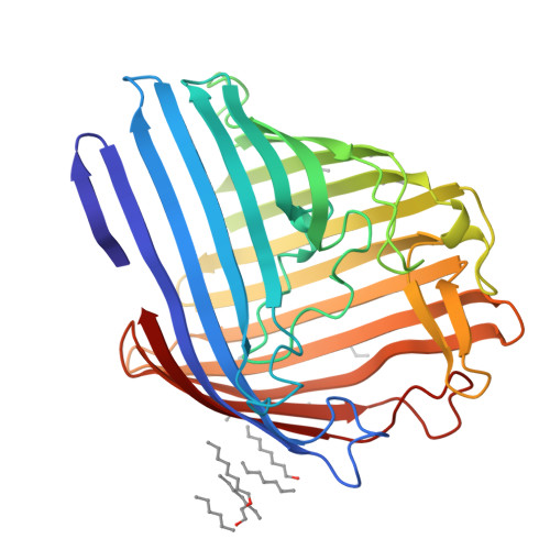
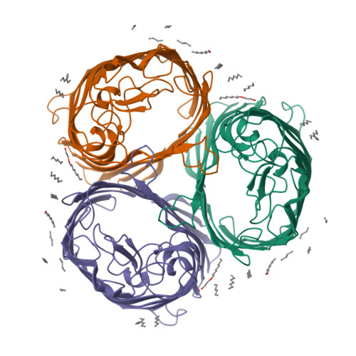
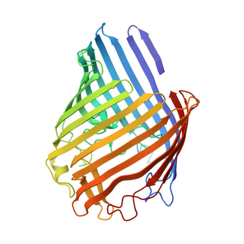
1MPF

STRUCTURAL AND FUNCTIONAL ALTERATIONS OF A COLICIN RESISTANT MUTANT OF OMPF-PORIN FROM ESCHERICHIA COLI


Experimental Data Snapshot

  • Method: X-RAY DIFFRACTION
  • Resolution: 3.00 Å
  • R-Value Work: 0.166 
  • R-Value Observed: 0.166 

wwPDB Validation   3D Report Full Report


This is version 1.3 of the entry. See complete history


Literature

Structural and functional alterations of a colicin-resistant mutant of OmpF porin from Escherichia coli.

Jeanteur, D.Schirmer, T.Fourel, D.Simonet, V.Rummel, G.Widmer, C.Rosenbusch, J.P.Pattus, F.Pages, J.M.

(1994) Proc Natl Acad Sci U S A 91: 10675-10679

  • DOI: https://doi.org/10.1073/pnas.91.22.10675
  • Primary Citation of Related Structures:  
    1MPF

  • PubMed Abstract: 

    A strain of Escherichia coli, selected on the basis of its resistance to colicin N, reveals distinct structural and functional alterations in unspecific OmpF porin. A single mutation [Gly-119-->Asp (G119D)] was identified in the internal loop L3 that contributes critically to the formation of the construction inside the lumen of the pore. X-ray structure analysis to a resolution of 3.0 A reveals a locally altered peptide backbone, with the side chain of residue Asp-119 protruding into the channel, causing the area of the constriction (7 x 11 A in the wild type) to be subdivided into two intercommunicating subcompartments of 3-4 A in diameter. The functional consequences of this structural modification consist of a reduction of the channel conductance by about one-third, of altered ion selectivity and voltage gating, and of a decrease of permeation rates of various sugars by factors of 2-12. The structural modification of the mutant protein affects neither the beta-barrel structure nor those regions of the molecule that are exposed at the cell surface. Considering the colicin resistance of the mutant, it is inferred that in vivo, colicin N traverses the outer membrane through the porin channel or that the dynamics of the exposed loops are affected in the mutant such that these may impede the binding of the toxin.


  • Organizational Affiliation

    European Molecular Biology Laboratory, Heidelberg, Germany.


Macromolecules
Find similar proteins by:  (by identity cutoff)  |  3D Structure
Entity ID: 1
MoleculeChains Sequence LengthOrganismDetailsImage
MATRIX PORIN OUTER MEMBRANE PROTEIN F340Escherichia coliMutation(s): 0 
Membrane Entity: Yes 
UniProt
Find proteins for P02931 (Escherichia coli (strain K12))
Explore P02931 
Go to UniProtKB:  P02931
Entity Groups  
Sequence Clusters30% Identity50% Identity70% Identity90% Identity95% Identity100% Identity
UniProt GroupP02931
Sequence Annotations
Expand
  • Reference Sequence
Small Molecules
Ligands 1 Unique
IDChains Name / Formula / InChI Key2D Diagram3D Interactions
C8E
Query on C8E

Download Ideal Coordinates CCD File 
B [auth A]
C [auth A]
D [auth A]
E [auth A]
F [auth A]
B [auth A],
C [auth A],
D [auth A],
E [auth A],
F [auth A],
G [auth A],
H [auth A],
I [auth A],
J [auth A],
K [auth A],
L [auth A],
M [auth A]
(HYDROXYETHYLOXY)TRI(ETHYLOXY)OCTANE
C16 H34 O5
FEOZZFHAVXYAMB-UHFFFAOYSA-N
Experimental Data & Validation

Experimental Data

  • Method: X-RAY DIFFRACTION
  • Resolution: 3.00 Å
  • R-Value Work: 0.166 
  • R-Value Observed: 0.166 
  • Space Group: P 3 2 1
Unit Cell:
Length ( Å )Angle ( ˚ )
a = 117.9α = 90
b = 117.9β = 90
c = 52.8γ = 120
Software Package:
Software NamePurpose
X-PLORmodel building
X-PLORrefinement
X-PLORphasing

Structure Validation

View Full Validation Report


Entry History 

Deposition Data

  • Released Date: 1995-02-07 
  • Deposition Author(s): Schirmer, T.

Revision History  (Full details and data files)

  • Version 1.0: 1995-02-07
    Type: Initial release
  • Version 1.1: 2008-03-24
    Changes: Version format compliance
  • Version 1.2: 2011-07-13
    Changes: Derived calculations, Version format compliance
  • Version 1.3: 2024-02-14
    Changes: Data collection, Database references, Derived calculations, Other