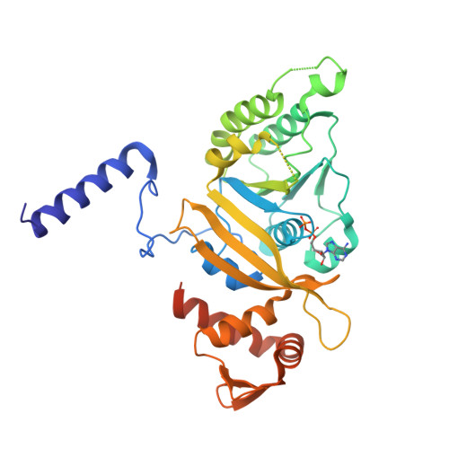
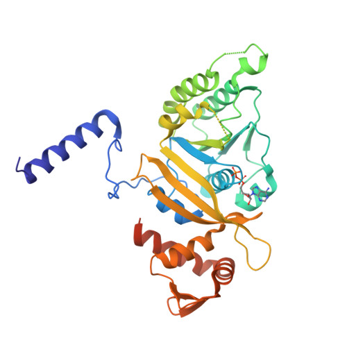
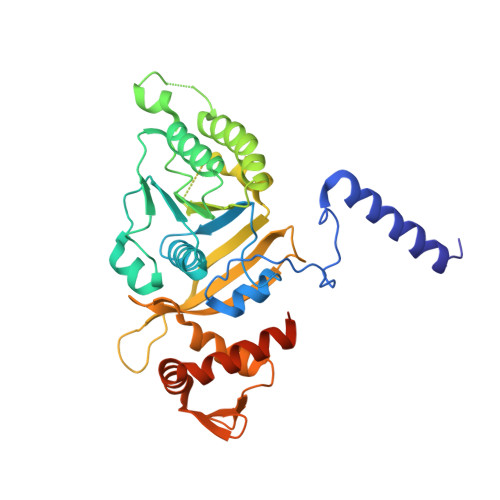
Structural studies on MtRecA-nucleotide complexes: Insights into DNA and nucleotide binding and the structural signature of NTP recognition
Datta, S., Ganesh, N., Chandra, N.R., Muniyappa, K., Vijayan, M.(2003) Proteins 50: 474-485
- PubMed: 12557189
- DOI: https://doi.org/10.1002/prot.10315
- Primary Citation of Related Structures:
1MO3, 1MO4, 1MO5, 1MO6 - PubMed Abstract:
RecA protein plays a crucial role in homologous recombination and repair of DNA. Central to all activities of RecA is its binding to Mg(+2)-ATP. The active form of the protein is a helical nucleoprotein filament containing the nucleotide cofactor and single-stranded DNA. The stability and structure of the helical nucleoprotein filament formed by RecA are modulated by nucleotide cofactors. Here we report crystal structures of a MtRecA-ADP complex, complexes with ATPgammaS in the presence and absence of magnesium as well as a complex with dATP and Mg+2. Comparison with the recently solved crystal structures of the apo form as well as a complex with ADP-AlF4 confirms an expansion of the P-loop region in MtRecA, compared to its homologue in Escherichia coli, correlating with the reduced affinity of MtRecA for ATP. The ligand bound structures reveal subtle variations in nucleotide conformations among different nucleotides that serve in maintaining the network of interactions crucial for nucleotide binding. The nucleotide binding site itself, however, remains relatively unchanged. The analysis also reveals that ATPgammaS rather than ADP-AlF4 is structurally a better mimic of ATP. From among the complexed structures, a definition for the two DNA-binding loops L1 and L2 has clearly emerged for the first time and provides a basis to understand DNA binding by RecA. The structural information obtained from these complexes correlates well with the extensive biochemical data on mutants available in the literature, contributing to an understanding of the role of individual residues in the nucleotide binding pocket, at the molecular level. Modeling studies on the mutants again point to the relative rigidity of the nucleotide binding site. Comparison with other NTP binding proteins reveals many commonalties in modes of binding by diverse members in the structural family, contributing to our understanding of the structural signature of NTP recognition.
Organizational Affiliation:
Molecular Biophysics Unit, Indian Institute of Science, Bangalore, India.


