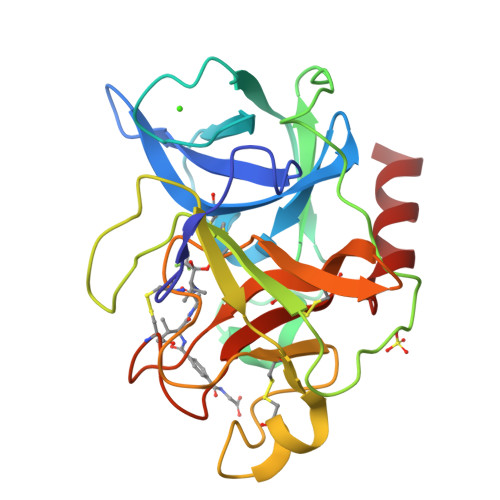
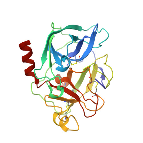
1MMJ

Porcine pancreatic elastase complexed with a potent peptidyl inhibitor, FR136706


Experimental Data Snapshot

  • Method: X-RAY DIFFRACTION
  • Resolution: 2.20 Å
  • R-Value Free: 0.241 
  • R-Value Work: 0.215 
  • R-Value Observed: 0.215 

wwPDB Validation   3D Report Full Report


Ligand Structure Quality Assessment 


This is version 1.4 of the entry. See complete history


Literature

True interaction mode of porcine pancreatic elastase with FR136706, a potent peptidyl inhibitor

Kinoshita, T.Nakanishi, I.Sato, A.Tada, T.

(2003) Bioorg Med Chem Lett 13: 21-24

  • DOI: https://doi.org/10.1016/s0960-894x(02)00852-1
  • Primary Citation of Related Structures:  
    1MMJ

  • PubMed Abstract: 

    The crystal structure of porcine pancreatic elastase (PPE) complexed with a potent peptidyl inhibitor, FR136706, was solved at 2.2A resolution. FR136706 fits snugly into the extended active site pocket. The benzene moiety of FR136706 induced dramatic movement of the side chain moiety of Arg217 and both moieties formed a pi-pi interaction, which has never been found previously in structures of PPE complexed with inhibitors. This novel interaction mode may lead to design of new types of inhibitors.


  • Organizational Affiliation

    Exploratory Research Laboratories, Fujisawa Pharmaceutical Co. Ltd., 5-2-3, Tokodai, Tsukuba, Ibaraki 300-2698, Japan. takayoshi_kinoshita@po.fujisawa.co.jp


Macromolecules
Find similar proteins by:  (by identity cutoff)  |  3D Structure
Entity ID: 1
MoleculeChains Sequence LengthOrganismDetailsImage
elastase 1A [auth N]240Sus scrofaMutation(s): 0 
EC: 3.4.21.36
UniProt
Find proteins for P00772 (Sus scrofa)
Explore P00772 
Go to UniProtKB:  P00772
Entity Groups  
Sequence Clusters30% Identity50% Identity70% Identity90% Identity95% Identity100% Identity
UniProt GroupP00772
Sequence Annotations
Expand
  • Reference Sequence
Small Molecules
Ligands 3 Unique
IDChains Name / Formula / InChI Key2D Diagram3D Interactions
FR1
Query on FR1

Download Ideal Coordinates CCD File 
D [auth N]2-[4-[[(S)-1-[[(S)-2-[[(RS)-3,3,3-TRIFLUORO-1-ISOPROPYL-2-OXOPROPYL]AMINOCARBONYL]PYRROLIDIN-1-YL-]CARBONYL]-2-METHYLPROPYL]AMINOCARBONYL]BENZOYLAMINO]ACETIC ACID
C26 H33 F3 N4 O7
MRTSIBBMOWLRPY-IHPCNDPISA-N
SO4
Query on SO4

Download Ideal Coordinates CCD File 
B [auth N]SULFATE ION
O4 S
QAOWNCQODCNURD-UHFFFAOYSA-L
CA
Query on CA

Download Ideal Coordinates CCD File 
C [auth N]CALCIUM ION
Ca
BHPQYMZQTOCNFJ-UHFFFAOYSA-N
Experimental Data & Validation

Experimental Data

  • Method: X-RAY DIFFRACTION
  • Resolution: 2.20 Å
  • R-Value Free: 0.241 
  • R-Value Work: 0.215 
  • R-Value Observed: 0.215 
  • Space Group: P 21 21 21
Unit Cell:
Length ( Å )Angle ( ˚ )
a = 51.29α = 90
b = 58.31β = 90
c = 75.52γ = 90
Software Package:
Software NamePurpose
AMoREphasing
X-PLORrefinement

Structure Validation

View Full Validation Report


Ligand Structure Quality Assessment 


Entry History 

Deposition Data

Revision History  (Full details and data files)

  • Version 1.0: 2002-12-23
    Type: Initial release
  • Version 1.1: 2008-04-28
    Changes: Version format compliance
  • Version 1.2: 2011-07-13
    Changes: Version format compliance
  • Version 1.3: 2017-10-11
    Changes: Refinement description
  • Version 1.4: 2024-11-20
    Changes: Data collection, Database references, Derived calculations, Structure summary