1MAM | pdb_00001mam

CRYSTAL STRUCTURE TO 2.45 A RESOLUTION OF A MONOCLONAL FAB SPECIFIC FOR THE BRUCELLA A CELL WALL POLYSACCHARIDE ANTIGEN


Experimental Data Snapshot

  • Method: X-RAY DIFFRACTION
  • Resolution: 2.45 Å
  • R-Value Work: 
    0.225 (DCC) 
  • R-Value Observed: 
    0.215 (Depositor) 

wwPDB Validation 3D Report Full Report

Validation slider image for 1MAM

This is version 1.4 of the entry. See complete history

Literature

Crystal structure to 2.45 A resolution of a monoclonal Fab specific for the Brucella A cell wall polysaccharide antigen.

Rose, D.R.Przybylska, M.To, R.J.Kayden, C.S.Oomen, R.P.Vorberg, E.Young, N.M.Bundle, D.R.

(1993) Protein Sci 2: 1106-1113

  • DOI: https://doi.org/10.1002/pro.5560020705
  • Primary Citation Related Structures: 
    1MAM

  • PubMed Abstract: 

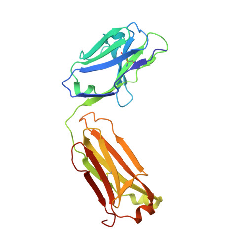
    The atomic structure of an antibody antigen-binding fragment (Fab) at 2.45 A resolution shows that polysaccharide antigen conformation and Fab structure dictated by combinatorial diversity and domain association are responsible for the fine specificity of the Brucella-specific antibody, YsT9.1. It discriminates the Brucella abortus A antigen from the nearly identical Brucella melitensis M antigen by forming a groove-type binding site, lined with tyrosine residues, that accommodates the rodlike A antigen but excludes the kinked structure of the M antigen, as envisioned by a model of the antigen built into the combining site. The variable-heavy (VH) and variable-light (VL) domains are derived from genes closely related to two used in previously solved structures, M603 and R19.9, respectively. These genes combine in YsT9.1 to form an antibody of totally different specificity. Comparison of this X-ray structure with a previously built model of the YsT9.1 combining site based on these homologies highlights the importance of VL:VH association as a determinant of specificity and suggests that small changes at the VL:VH interface, unanticipated in modeling, may cause significant modulation of binding-site properties.


  • Organizational Affiliation
    • Ontario Cancer Institute, University of Toronto, Canada.

Macromolecule Content 

  • Total Structure Weight: 46.93 kDa 
  • Atom Count: 3,296 
  • Modeled Residue Count: 431 
  • Deposited Residue Count: 431 
  • Unique protein chains: 2

Macromolecules

Find similar proteins by:|  3D Structure
Entity ID: 1
MoleculeChains  Sequence LengthOrganismDetailsImage
IGG2B-KAPPA YST9.1 FAB (LIGHT CHAIN)A [auth L]214Mus musculusMutation(s): 0 
Find similar proteins by:|  3D Structure
Entity ID: 2
MoleculeChains  Sequence LengthOrganismDetailsImage
IGG2B-KAPPA YST9.1 FAB (HEAVY CHAIN)B [auth H]217Mus musculusMutation(s): 0 
UniProt
Find proteins for P01867 (Mus musculus)
Explore P01867 
Go to UniProtKB:  P01867
Entity Groups
Sequence Clusters30% Identity50% Identity70% Identity90% Identity95% Identity100% Identity
UniProt GroupP01867
Sequence Annotations
Expand
Reference Sequence

Experimental Data & Validation

Experimental Data

  • Method: X-RAY DIFFRACTION
  • Resolution: 2.45 Å
  • R-Value Work:  0.225 (DCC) 
  • R-Value Observed: 0.215 (Depositor) 
Space Group: P 21 21 21
Unit Cell:
Length ( Å )Angle ( ˚ )
a = 75.73α = 90
b = 126.82β = 90
c = 46.82γ = 90
Software Package:
Software NamePurpose
PROLSQrefinement

Structure Validation

View Full Validation Report


Entry History 

Deposition Data

  • Released Date: 1993-10-31 
  • Deposition Author(s): Rose, D.R.

Revision History  (Full details and data files)

  • Version 1.0: 1993-10-31
    Type: Initial release
  • Version 1.1: 2008-03-24
    Changes: Version format compliance
  • Version 1.2: 2011-07-13
    Changes: Version format compliance
  • Version 1.3: 2024-06-05
    Changes: Data collection, Database references, Other
  • Version 1.4: 2024-10-23
    Changes: Structure summary