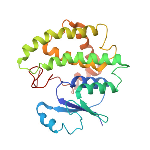
1M9B

Crystal structure of the 26 kDa glutathione S-transferase from Schistosoma japonicum complexed with gamma-glutamyl[S-(2-iodobenzyl)cysteinyl]glycine


Experimental Data Snapshot

  • Method: X-RAY DIFFRACTION
  • Resolution: 2.60 Å
  • R-Value Free: 0.275 
  • R-Value Work: 0.231 

Starting Model: experimental
View more details

wwPDB Validation   3D Report Full Report


Ligand Structure Quality Assessment 


This is version 1.3 of the entry. See complete history


Literature

Characterization of the electrophile binding site and substrate binding mode of the 26-kDa glutathione S-transferase from Schistosoma japonicum

Cardoso, R.M.F.Daniels, D.S.Bruns, C.M.Tainer, J.A.

(2003) Proteins 51: 137-146

  • DOI: https://doi.org/10.1002/prot.10345
  • Primary Citation of Related Structures:  
    1M99, 1M9A, 1M9B

  • PubMed Abstract: 

    The 26-kDa glutathione S-transferase from Schistosoma japonicum (Sj26GST), a helminth worm that causes schistosomiasis, catalyzes the conjugation of glutathione with toxic secondary products of membrane lipid peroxidation. Crystal structures of Sj26GST in complex with glutathione sulfonate (Sj26GSTSLF), S-hexyl glutathione (Sj26GSTHEX), and S-2-iodobenzyl glutathione (Sj26GSTIBZ) allow characterization of the electrophile binding site (H site) of Sj26GST. The S-hexyl and S-2-iodobenzyl moieties of these product analogs bind in a pocket defined by side-chains from the beta1-alpha1 loop (Tyr7, Trp8, Ile10, Gly12, Leu13), helix alpha4 (Arg103, Tyr104, Ser107, Tyr111), and the C-terminal coil (Gln204, Gly205, Trp206, Gln207). Changes in the Ser107 and Gln204 dihedral angles make the H site more hydrophobic in the Sj26GSTHEX complex relative to the ligand-free structure. These structures, together with docking studies, indicate a possible binding mode of Sj26GST to its physiologic substrates 4-hydroxynon-2-enal (4HNE), trans-non-2-enal (NE), and ethacrynic acid (EA). In this binding mode, hydrogen bonds of Tyr111 and Gln207 to the carbonyl oxygen atoms of 4HNE, NE, and EA could orient the substrates and enhance their electrophilicity to promote conjugation with glutathione.


  • Organizational Affiliation

    Skaggs Institute for Chemical Biology, The Scripps Research Institute, La Jolla, CA 92037, USA.


Macromolecules
Find similar proteins by:  (by identity cutoff)  |  3D Structure
Entity ID: 1
MoleculeChains Sequence LengthOrganismDetailsImage
Glutathione S-Transferase 26 kDa218Schistosoma japonicumMutation(s): 0 
EC: 2.5.1.18
UniProt
Find proteins for P08515 (Schistosoma japonicum)
Explore P08515 
Go to UniProtKB:  P08515
Entity Groups  
Sequence Clusters30% Identity50% Identity70% Identity90% Identity95% Identity100% Identity
UniProt GroupP08515
Sequence Annotations
Expand
  • Reference Sequence
Small Molecules
Ligands 1 Unique
IDChains Name / Formula / InChI Key2D Diagram3D Interactions
IBG
Query on IBG

Download Ideal Coordinates CCD File 
B [auth A]GAMMA-GLUTAMYL[S-(2-IODOBENZYL)CYSTEINYL]GLYCINE
C17 H22 I N3 O6 S
QFYJAEOZTBVJQM-STQMWFEESA-N
Experimental Data & Validation

Experimental Data

  • Method: X-RAY DIFFRACTION
  • Resolution: 2.60 Å
  • R-Value Free: 0.275 
  • R-Value Work: 0.231 
  • Space Group: P 63 2 2
Unit Cell:
Length ( Å )Angle ( ˚ )
a = 116.567α = 90
b = 116.567β = 90
c = 78.748γ = 120
Software Package:
Software NamePurpose
DENZOdata reduction
SCALEPACKdata scaling
AMoREphasing
CNSrefinement

Structure Validation

View Full Validation Report


Ligand Structure Quality Assessment 


Entry History 

Deposition Data

Revision History  (Full details and data files)

  • Version 1.0: 2003-03-04
    Type: Initial release
  • Version 1.1: 2008-04-28
    Changes: Version format compliance
  • Version 1.2: 2011-07-13
    Changes: Derived calculations, Version format compliance
  • Version 1.3: 2024-02-14
    Changes: Data collection, Database references, Derived calculations, Refinement description