1M73 | pdb_00001m73

CRYSTAL STRUCTURE OF HUMAN PNP AT 2.3A RESOLUTION


Experimental Data Snapshot

  • Method: X-RAY DIFFRACTION
  • Resolution: 2.30 Å
  • R-Value Free: 
    0.222 (Depositor), 0.205 (DCC) 
  • R-Value Work: 
    0.205 (Depositor), 0.192 (DCC) 
  • R-Value Observed: 
    0.239 (Depositor) 

Starting Model: experimental
View more details

wwPDB Validation 3D Report Full Report

Validation slider image for 1M73

This is version 1.3 of the entry. See complete history

Literature

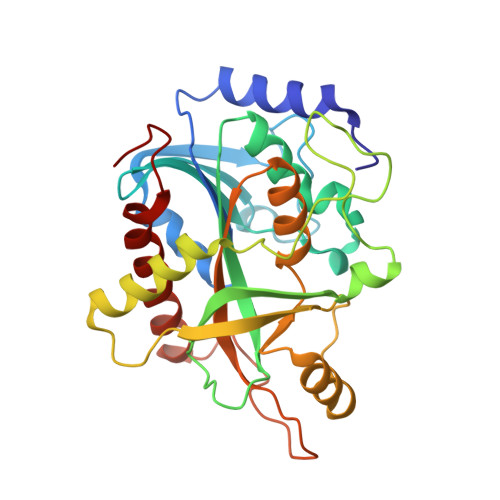
Crystal structure of human purine nucleoside phosphorylase at 2.3A resolution.

de Azevedo, W.F.Canduri, F.dos Santos, D.M.Silva, R.G.de Oliveira, J.S.de Carvalho, L.P.Basso, L.A.Mendes, M.A.Palma, M.S.Santos, D.S.

(2003) Biochem Biophys Res Commun 308: 545-552

  • DOI: https://doi.org/10.1016/s0006-291x(03)01431-1
  • Primary Citation Related Structures: 
    1M73

  • PubMed Abstract: 

    Purine nucleoside phosphorylase (PNP) catalyzes the phosphorolysis of the N-ribosidic bonds of purine nucleosides and deoxynucleosides. In human, PNP is the only route for degradation of deoxyguanosine and genetic deficiency of this enzyme leads to profound T-cell mediated immunosuppression. PNP is therefore a target for inhibitor development aiming at T-cell immune response modulation and its low resolution structure has been used for drug design. Here we report the structure of human PNP solved to 2.3A resolution using synchrotron radiation and cryocrystallographic techniques. This structure allowed a more precise analysis of the active site, generating a more reliable model for substrate binding. The higher resolution data allowed the identification of water molecules in the active site, which suggests binding partners for potential ligands. Furthermore, the present structure may be used in the new structure-based design of PNP inhibitors.


  • Organizational Affiliation
    • Departamento de Física, UNESP, São José do Rio Preto, SP, Brazil. walterfa@df.ibilce.unesp.br

Macromolecule Content 

  • Total Structure Weight: 32.34 kDa 
  • Atom Count: 2,334 
  • Modeled Residue Count: 288 
  • Deposited Residue Count: 288 
  • Unique protein chains: 1

Macromolecules

Find similar proteins by:|  3D Structure
Entity ID: 1
MoleculeChains  Sequence LengthOrganismDetailsImage
PURINE NUCLEOSIDE PHOSPHORYLASEA [auth E]288Homo sapiensMutation(s): 0 
EC: 2.4.2.1
UniProt & NIH Common Fund Data Resources
Find proteins for P00491 (Homo sapiens)
Explore P00491 
Go to UniProtKB:  P00491
PHAROS:  P00491
GTEx:  ENSG00000198805 
Entity Groups
Sequence Clusters30% Identity50% Identity70% Identity90% Identity95% Identity100% Identity
UniProt GroupP00491
Sequence Annotations
Expand
Reference Sequence

Experimental Data & Validation

Experimental Data

  • Method: X-RAY DIFFRACTION
  • Resolution: 2.30 Å
  • R-Value Free:  0.222 (Depositor), 0.205 (DCC) 
  • R-Value Work:  0.205 (Depositor), 0.192 (DCC) 
  • R-Value Observed: 0.239 (Depositor) 
Space Group: H 3 2
Unit Cell:
Length ( Å )Angle ( ˚ )
a = 141.63α = 90
b = 141.63β = 90
c = 163.16γ = 120
Software Package:
Software NamePurpose
DENZOdata reduction
SCALEPACKdata scaling
AMoREphasing
X-PLORrefinement

Structure Validation

View Full Validation Report


Entry History 

Revision History  (Full details and data files)

  • Version 1.0: 2003-09-16
    Type: Initial release
  • Version 1.1: 2008-04-28
    Changes: Version format compliance
  • Version 1.2: 2011-07-13
    Changes: Derived calculations, Version format compliance
  • Version 1.3: 2023-10-25
    Changes: Data collection, Database references, Derived calculations, Refinement description