1M6V | pdb_00001m6v

Crystal Structure of the G359F (small subunit) Point Mutant of Carbamoyl Phosphate Synthetase


Experimental Data Snapshot

  • Method: X-RAY DIFFRACTION
  • Resolution: 2.10 Å
  • R-Value Free: 
    0.245 (Depositor), 0.176 (DCC) 
  • R-Value Work: 
    0.174 (Depositor), 0.176 (DCC) 
  • R-Value Observed: 
    0.180 (Depositor) 

Starting Model: experimental
View more details

wwPDB Validation 3D Report Full Report

Validation slider image for 1M6V

Ligand Structure Quality Assessment 


This is version 1.5 of the entry. See complete history

Literature

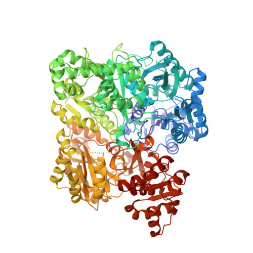
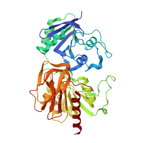
Carbamoyl-phosphate synthetase. Creation of an escape route for ammonia

Thoden, J.B.Huang, X.Raushel, F.M.Holden, H.M.

(2002) J Biological Chem 277: 39722-39727

  • DOI: https://doi.org/10.1074/jbc.M206915200
  • Primary Citation Related Structures: 
    1M6V

  • PubMed Abstract: 

    Carbamoyl-phosphate synthetase catalyzes the production of carbamoyl phosphate through a reaction mechanism requiring one molecule of bicarbonate, two molecules of MgATP, and one molecule of glutamine. The enzyme from Escherichia coli is composed of two polypeptide chains. The smaller of these belongs to the Class I amidotransferase superfamily and contains all of the necessary amino acid side chains required for the hydrolysis of glutamine to glutamate and ammonia. Two homologous domains from the larger subunit adopt conformations that are characteristic for members of the ATP-grasp superfamily. Each of these ATP-grasp domains contains an active site responsible for binding one molecule of MgATP. High resolution x-ray crystallographic analyses have shown that, remarkably, the three active sites in the E. coli enzyme are connected by a molecular tunnel of approximately 100 A in total length. Here we describe the high resolution x-ray crystallographic structure of the G359F (small subunit) mutant protein of carbamoyl phosphate synthetase. This residue was initially targeted for study because it resides within the interior wall of the molecular tunnel leading from the active site of the small subunit to the first active site of the large subunit. It was anticipated that a mutation to the larger residue would "clog" the ammonia tunnel and impede the delivery of ammonia from its site of production to the site of utilization. In fact, the G359F substitution resulted in a complete change in the conformation of the loop delineated by Glu-355 to Ala-364, thereby providing an "escape" route for the ammonia intermediate directly to the bulk solvent. The substitution also effected the disposition of several key catalytic amino acid side chains in the small subunit active site.


  • Organizational Affiliation
    • Department of Biochemistry, University of Wisconsin, Madison, Wisconsin, 53706-1544, USA.

Macromolecule Content 

  • Total Structure Weight: 645.73 kDa 
  • Atom Count: 48,206 
  • Modeled Residue Count: 5,736 
  • Deposited Residue Count: 5,820 
  • Unique protein chains: 2

Macromolecules

Find similar proteins by:|  3D Structure
Entity ID: 1
MoleculeChains  Sequence LengthOrganismDetailsImage
carbamoyl phosphate synthetase large chain
A, C, E, G
1,073Escherichia coliMutation(s): 0 
Gene Names: CARB
EC: 6.3.5.5 (PDB Primary Data), 6.3.4.16 (UniProt)
UniProt
Find proteins for P00968 (Escherichia coli (strain K12))
Explore P00968 
Go to UniProtKB:  P00968
Entity Groups
Sequence Clusters30% Identity50% Identity70% Identity90% Identity95% Identity100% Identity
UniProt GroupP00968
Sequence Annotations
Expand
Reference Sequence
Find similar proteins by:|  3D Structure
Entity ID: 2
MoleculeChains  Sequence LengthOrganismDetailsImage
carbamoyl-phosphate synthetase small chain
B, D, F, H
382Escherichia coliMutation(s): 1 
Gene Names: CARA
EC: 6.3.5.5
UniProt
Find proteins for P0A6F1 (Escherichia coli (strain K12))
Explore P0A6F1 
Go to UniProtKB:  P0A6F1
Entity Groups
Sequence Clusters30% Identity50% Identity70% Identity90% Identity95% Identity100% Identity
UniProt GroupP0A6F1
Sequence Annotations
Expand
Reference Sequence

Small Molecules

Ligands 7 Unique
IDChains Name / Formula / InChI Key2D Diagram3D Interactions
ADP

Query on ADP


Download:Ideal Coordinates CCD File
LC [auth G]
MC [auth G]
PB [auth E]
QB [auth E]
TA [auth C]
LC [auth G],
MC [auth G],
PB [auth E],
QB [auth E],
TA [auth C],
UA [auth C],
W [auth A],
X [auth A]
ADENOSINE-5'-DIPHOSPHATE
C10 H15 N5 O10 P2
XTWYTFMLZFPYCI-KQYNXXCUSA-N
ORN

Query on ORN


Download:Ideal Coordinates CCD File
NC [auth G],
RB [auth E],
VA [auth C],
Y [auth A]
L-ornithine
C5 H12 N2 O2
AHLPHDHHMVZTML-BYPYZUCNSA-N
NET

Query on NET


Download:Ideal Coordinates CCD File
OC [auth G],
SB [auth E],
WA [auth C],
Z [auth A]
TETRAETHYLAMMONIUM ION
C8 H20 N
CBXCPBUEXACCNR-UHFFFAOYSA-N
PO4

Query on PO4


Download:Ideal Coordinates CCD File
KC [auth G],
OB [auth E],
SA [auth C],
V [auth A]
PHOSPHATE ION
O4 P
NBIIXXVUZAFLBC-UHFFFAOYSA-K
MN

Query on MN


Download:Ideal Coordinates CCD File
AB [auth E]
CA [auth C]
DA [auth C]
DB [auth E]
GA [auth C]
AB [auth E],
CA [auth C],
DA [auth C],
DB [auth E],
GA [auth C],
I [auth A],
J [auth A],
M [auth A],
VB [auth G],
WB [auth G],
ZA [auth E],
ZB [auth G]
MANGANESE (II) ION
Mn
WAEMQWOKJMHJLA-UHFFFAOYSA-N
K

Query on K


Download:Ideal Coordinates CCD File
AA [auth B]
AC [auth G]
BB [auth E]
BC [auth G]
CB [auth E]
AA [auth B],
AC [auth G],
BB [auth E],
BC [auth G],
CB [auth E],
CC [auth G],
DC [auth G],
EA [auth C],
EB [auth E],
FA [auth C],
FB [auth E],
GB [auth E],
HA [auth C],
HB [auth E],
IA [auth C],
JA [auth C],
K [auth A],
KA [auth C],
L [auth A],
N [auth A],
O [auth A],
P [auth A],
TB [auth F],
XA [auth D],
XB [auth G],
YB [auth G]
POTASSIUM ION
K
NPYPAHLBTDXSSS-UHFFFAOYSA-N
CL

Query on CL


Download:Ideal Coordinates CCD File
BA [auth B]
EC [auth G]
FC [auth G]
GC [auth G]
HC [auth G]
BA [auth B],
EC [auth G],
FC [auth G],
GC [auth G],
HC [auth G],
IB [auth E],
IC [auth G],
JB [auth E],
JC [auth G],
KB [auth E],
LA [auth C],
LB [auth E],
MA [auth C],
MB [auth E],
NA [auth C],
NB [auth E],
OA [auth C],
PA [auth C],
PC [auth H],
Q [auth A],
QA [auth C],
R [auth A],
RA [auth C],
S [auth A],
T [auth A],
U [auth A],
UB [auth F],
YA [auth D]
CHLORIDE ION
Cl
VEXZGXHMUGYJMC-UHFFFAOYSA-M

Experimental Data & Validation

Experimental Data

  • Method: X-RAY DIFFRACTION
  • Resolution: 2.10 Å
  • R-Value Free:  0.245 (Depositor), 0.176 (DCC) 
  • R-Value Work:  0.174 (Depositor), 0.176 (DCC) 
  • R-Value Observed: 0.180 (Depositor) 
Space Group: P 21 21 21
Unit Cell:
Length ( Å )Angle ( ˚ )
a = 151.5α = 90
b = 164.2β = 90
c = 331.5γ = 90
Software Package:
Software NamePurpose
d*TREKdata scaling
HKL-2000data reduction
TNTrefinement
d*TREKdata reduction
HKL-2000data scaling
TNTphasing

Structure Validation

View Full Validation Report


Ligand Structure Quality Assessment 


Entry History 

Deposition Data

Revision History  (Full details and data files)

  • Version 1.0: 2002-07-31
    Type: Initial release
  • Version 1.1: 2008-04-28
    Changes: Version format compliance
  • Version 1.2: 2011-07-13
    Changes: Non-polymer description, Version format compliance
  • Version 1.3: 2019-11-20
    Changes: Advisory, Derived calculations
  • Version 1.4: 2021-10-27
    Changes: Database references, Derived calculations
  • Version 1.5: 2024-02-14
    Changes: Data collection, Refinement description