1M6J | pdb_00001m6j

CRYSTAL STRUCTURE OF TRIOSEPHOSPHATE ISOMERASE FROM ENTAMOEBA HISTOLYTICA


Experimental Data Snapshot

  • Method: X-RAY DIFFRACTION
  • Resolution: 1.50 Å
  • R-Value Free: 
    0.206 (Depositor), 0.210 (DCC) 
  • R-Value Work: 
    0.184 (Depositor), 0.189 (DCC) 
  • R-Value Observed: 
    0.184 (Depositor) 

Starting Model: experimental
View more details

wwPDB Validation 3D Report Full Report

Validation slider image for 1M6J

This is version 1.3 of the entry. See complete history

Literature

Structure and Inactivation of Triosephosphate Isomerase from Entamoeba histolytica

Rodriguez-Romero, A.Hernandez-Santoyo, A.Del Pozo-Yauner, L.Kornhauser, A.Fernandez-Velasco, D.A.

(2002) J Mol Biology 322: 669-675

  • DOI: https://doi.org/10.1016/s0022-2836(02)00809-4
  • Primary Citation Related Structures: 
    1M6J

  • PubMed Abstract: 

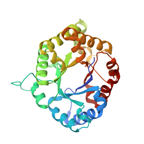
    Triosephosphate isomerase (TIM) has been proposed as a target for drug design. TIMs from several parasites have a cysteine residue at the dimer interface, whose derivatization with thiol-specific reagents induces enzyme inactivation and aggregation. TIMs lacking this residue, such as human TIM, are less affected. TIM from Entamoeba histolytica (EhTIM) has the interface cysteine residue and presents more than ten insertions when compared with the enzyme from other pathogens. To gain further insight into the role that interface residues play in the stability and reactivity of these enzymes, we determined the high-resolution structure and characterized the effect of methylmethane thiosulfonate (MMTS) on the activity and conformational properties of EhTIM. The structure of this enzyme was determined at 1.5A resolution using molecular replacement, observing that the dimer is not symmetric. EhTIM is completely inactivated by MMTS, and dissociated into stable monomers that possess considerable secondary structure. Structural and spectroscopic analysis of EhTIM and comparison with TIMs from other pathogens reveal that conformational rearrangements of the interface after dissociation, as well as intramonomeric contacts formed by the inserted residues, may contribute to the unusual stability of the derivatized EhTIM monomer.


  • Organizational Affiliation
    • Laboratorio Universitario de Estructura de Proteínas and Departamento de Bioquímica, Instituto de Química, Universidad Nacional Autónoma de México, DF, Mexico. adela@servidor.unam.mx

Macromolecule Content 

  • Total Structure Weight: 55.93 kDa 
  • Atom Count: 4,806 
  • Modeled Residue Count: 520 
  • Deposited Residue Count: 522 
  • Unique protein chains: 1

Macromolecules

Find similar proteins by:|  3D Structure
Entity ID: 1
MoleculeChains  Sequence LengthOrganismDetailsImage
Triosephosphate Isomerase
A, B
261Entamoeba histolyticaMutation(s): 0 
EC: 5.3.1.1
UniProt
Find proteins for O02611 (Entamoeba histolytica (strain ATCC 30459 / HM-1:IMSS / ABRM))
Explore O02611 
Go to UniProtKB:  O02611
Entity Groups
Sequence Clusters30% Identity50% Identity70% Identity90% Identity95% Identity100% Identity
UniProt GroupO02611
Sequence Annotations
Expand
Reference Sequence

Experimental Data & Validation

Experimental Data

  • Method: X-RAY DIFFRACTION
  • Resolution: 1.50 Å
  • R-Value Free:  0.206 (Depositor), 0.210 (DCC) 
  • R-Value Work:  0.184 (Depositor), 0.189 (DCC) 
  • R-Value Observed: 0.184 (Depositor) 
Space Group: P 21 21 2
Unit Cell:
Length ( Å )Angle ( ˚ )
a = 85.729α = 90
b = 119.947β = 90
c = 50.392γ = 90
Software Package:
Software NamePurpose
CNSrefinement
MOSFLMdata reduction
CCP4data scaling
CNSphasing

Structure Validation

View Full Validation Report


Entry History 

Deposition Data

Revision History  (Full details and data files)

  • Version 1.0: 2002-10-12
    Type: Initial release
  • Version 1.1: 2008-04-28
    Changes: Version format compliance
  • Version 1.2: 2011-07-13
    Changes: Version format compliance
  • Version 1.3: 2024-02-14
    Changes: Data collection, Database references, Refinement description