1M55 | pdb_00001m55

Catalytic domain of the Adeno Associated Virus type 5 Rep protein


Experimental Data Snapshot

  • Method: X-RAY DIFFRACTION
  • Resolution: 1.40 Å
  • R-Value Free: 
    0.196 (Depositor) 
  • R-Value Work: 
    0.178 (Depositor) 

wwPDB Validation 3D Report Full Report

Validation slider image for 1M55

This is version 1.3 of the entry. See complete history

Literature

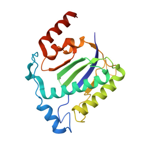
Structural unity among viral origin binding proteins: crystal structure of the nuclease domain of adeno-associated virus Rep.

Hickman, A.B.Ronning, D.R.Kotin, R.M.Dyda, F.

(2002) Mol Cell 10: 327-337

  • DOI: https://doi.org/10.1016/s1097-2765(02)00592-0
  • Primary Citation Related Structures: 
    1M55

  • PubMed Abstract: 

    Adeno-associated virus (AAV), unique among animal viruses in its ability to integrate into a specific chromosomal location, is a promising vector for human gene therapy. AAV Replication (Rep) protein is essential for viral replication and integration, and its amino terminal domain possesses site-specific DNA binding and endonuclease activities required for replication initiation and integration. This domain displays a novel endonuclease fold and demonstrates an unexpected structural relationship to other viral origin binding proteins such as the papillomavirus E1 protein and the SV40 T antigen. The active site, located at the bottom of a positively charged cleft, is formed by the spatial convergence of a divalent metal ion and two conserved sequence motifs that define the rolling circle replication superfamily.


  • Organizational Affiliation
    • Laboratory of Molecular Biology, National Institute of Diabetes and Digestive and Kidney Diseases, National Institutes of Health, Bethesda, MD 20892, USA. ahickman@helix.nih.gov

Macromolecule Content 

  • Total Structure Weight: 46.02 kDa 
  • Atom Count: 3,843 
  • Modeled Residue Count: 386 
  • Deposited Residue Count: 394 
  • Unique protein chains: 1

Macromolecules

Find similar proteins by:|  3D Structure
Entity ID: 1
MoleculeChains  Sequence LengthOrganismDetailsImage
Rep protein
A, B
197adeno-associated virus 5Mutation(s): 0 
UniProt
Find proteins for Q9YJC1 (adeno-associated virus 5)
Explore Q9YJC1 
Go to UniProtKB:  Q9YJC1
Entity Groups
Sequence Clusters30% Identity50% Identity70% Identity90% Identity95% Identity100% Identity
UniProt GroupQ9YJC1
Sequence Annotations
Expand
Reference Sequence

Experimental Data & Validation

Experimental Data

  • Method: X-RAY DIFFRACTION
  • Resolution: 1.40 Å
  • R-Value Free:  0.196 (Depositor) 
  • R-Value Work:  0.178 (Depositor) 
Space Group: P 1 21 1
Unit Cell:
Length ( Å )Angle ( ˚ )
a = 42.888α = 90
b = 73.455β = 94.491
c = 65.56γ = 90
Software Package:
Software NamePurpose
PHASESphasing
X-PLORrefinement
DENZOdata reduction
SCALEPACKdata scaling

Structure Validation

View Full Validation Report


Entry History 

Deposition Data

Revision History  (Full details and data files)

  • Version 1.0: 2002-08-28
    Type: Initial release
  • Version 1.1: 2008-04-28
    Changes: Version format compliance
  • Version 1.2: 2011-07-13
    Changes: Version format compliance
  • Version 1.3: 2024-02-14
    Changes: Data collection, Database references, Derived calculations