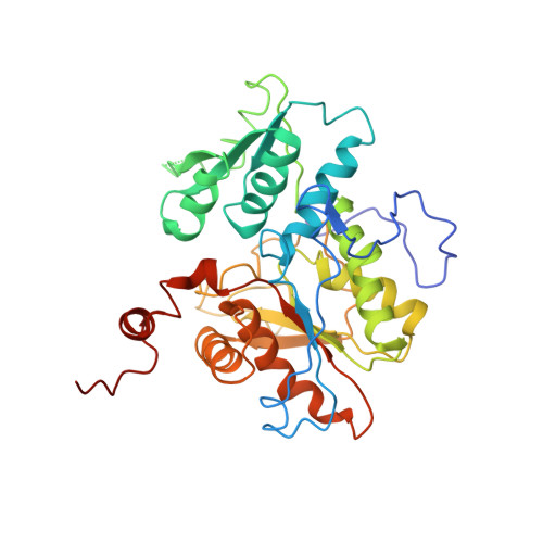
1M54

CYSTATHIONINE-BETA SYNTHASE: REDUCED VICINAL THIOLS


Experimental Data Snapshot

  • Method: X-RAY DIFFRACTION
  • Resolution: 2.90 Å
  • R-Value Free: 0.365 
  • R-Value Work: 0.271 

wwPDB Validation   3D Report Full Report


Ligand Structure Quality Assessment 


This is version 1.3 of the entry. See complete history


Literature

HUMAN CYSTATHIONINE BETA-SYNTHASE IS A HEME SENSOR PROTEIN. EVIDENCE THAT THE REDOX SENSOR IS HEME AND NOT THE VICINAL CYSTEINES IN THE CXXC MOTIF SEEN IN THE CRYSTAL STRUCTURE OF THE TRUNCATED ENZYME

Taoka, S.Lepore, B.W.Kabil, O.Ojha, S.Ringe, D.Banerjee, R.

(2002) Biochemistry 41: 10454-10461

  • DOI: https://doi.org/10.1021/bi026052d
  • Primary Citation of Related Structures:  
    1M54

  • PubMed Abstract: 

    Elevated levels of homocysteine, a sulfur-containing amino acid, are correlated with increased risk for cardiovascular diseases and Alzheimers disease and with neural tube defects. The only route for the catabolic removal of homocysteine in mammals begins with the pyridoxal phosphate- (PLP-) dependent beta-replacement reaction catalyzed by cystathionine beta-synthase. The enzyme has a b-type heme with unusual spectroscopic properties but as yet unknown function. The human enzyme has a modular organization and can be cleaved into an N-terminal catalytic core, which retains both the heme and PLP-binding sites and is highly active, and a C-terminal regulatory domain, where the allosteric activator S-adenosylmethionine is presumed to bind. Studies with the isolated recombinant enzyme and in transformed human liver cells indicate that the enzyme is approximately 2-fold more active under oxidizing conditions. In addition to heme, the enzyme contains a CXXC oxidoreductase motif that could, in principle, be involved in redox sensing. In this study, we have examined the role of heme versus the vicinal thiols in modulating the redox responsiveness of the enzyme. Deletion of the heme domain leads to loss of redox sensitivity. In contrast, substitution of either cysteine with a non-redox-active amino acid does not affect the responsiveness of the enzyme to reductants. We also report the crystal structure of the catalytic core of the enzyme in which the vicinal cysteines are reduced without any discernible differences in the remainder of the protein. The structure of the catalytic core is compared to those of other members of the fold II family of PLP-dependent enzymes and provides insights into active site residues that may be important in interacting with the substrates and intermediates.


  • Organizational Affiliation

    Biochemistry Department, University of Nebraska, Lincoln, Nebraska 68588-0664, USA.


Macromolecules
Find similar proteins by:  (by identity cutoff)  |  3D Structure
Entity ID: 1
MoleculeChains Sequence LengthOrganismDetailsImage
CYSTATHIONINE BETA-SYNTHASE
A, B, C, D, E
A, B, C, D, E, F
363Homo sapiensMutation(s): 1 
Gene Names: CBS
EC: 4.2.1.22
UniProt & NIH Common Fund Data Resources
Find proteins for P35520 (Homo sapiens)
Explore P35520 
Go to UniProtKB:  P35520
PHAROS:  P35520
GTEx:  ENSG00000160200 
Entity Groups  
Sequence Clusters30% Identity50% Identity70% Identity90% Identity95% Identity100% Identity
UniProt GroupP35520
Sequence Annotations
Expand
  • Reference Sequence
Small Molecules
Ligands 2 Unique
IDChains Name / Formula / InChI Key2D Diagram3D Interactions
HEM
Query on HEM

Download Ideal Coordinates CCD File 
H [auth A]
J [auth B]
L [auth C]
N [auth D]
P [auth E]
H [auth A],
J [auth B],
L [auth C],
N [auth D],
P [auth E],
R [auth F]
PROTOPORPHYRIN IX CONTAINING FE
C34 H32 Fe N4 O4
KABFMIBPWCXCRK-RGGAHWMASA-L
PLP
Query on PLP

Download Ideal Coordinates CCD File 
G [auth A]
I [auth B]
K [auth C]
M [auth D]
O [auth E]
G [auth A],
I [auth B],
K [auth C],
M [auth D],
O [auth E],
Q [auth F]
PYRIDOXAL-5'-PHOSPHATE
C8 H10 N O6 P
NGVDGCNFYWLIFO-UHFFFAOYSA-N
Experimental Data & Validation

Experimental Data

  • Method: X-RAY DIFFRACTION
  • Resolution: 2.90 Å
  • R-Value Free: 0.365 
  • R-Value Work: 0.271 
  • Space Group: P 1
Unit Cell:
Length ( Å )Angle ( ˚ )
a = 85.74α = 102.01
b = 85.626β = 101.53
c = 97.082γ = 112.6
Software Package:
Software NamePurpose
AMoREphasing
CNSrefinement
DENZOdata reduction
SCALEPACKdata scaling

Structure Validation

View Full Validation Report


Ligand Structure Quality Assessment 


Entry History 

Deposition Data

Revision History  (Full details and data files)

  • Version 1.0: 2002-08-14
    Type: Initial release
  • Version 1.1: 2008-04-28
    Changes: Version format compliance
  • Version 1.2: 2011-07-13
    Changes: Non-polymer description, Version format compliance
  • Version 1.3: 2021-10-27
    Changes: Database references, Derived calculations