1M4Y | pdb_00001m4y

Crystal structure of HslV from Thermotoga maritima


Experimental Data Snapshot

  • Method: X-RAY DIFFRACTION
  • Resolution: 2.10 Å
  • R-Value Free: 
    0.229 (Depositor), 0.229 (DCC) 
  • R-Value Work: 
    0.192 (Depositor), 0.192 (DCC) 

wwPDB Validation 3D Report Full Report

Validation slider image for 1M4Y

This is version 1.3 of the entry. See complete history

Literature

Isolation and characterization of the prokaryotic proteasome homolog HslVU (ClpQY) from Thermotoga maritima and the crystal structure of HslV.

Song, H.K.Bochtler, M.Azim, M.K.Hartmann, C.Huber, R.Ramachandran, R.

(2003) Biophys Chem 100: 437-452

  • DOI: https://doi.org/10.1016/s0301-4622(02)00297-1
  • Primary Citation Related Structures: 
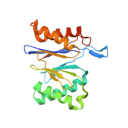
    1M4Y

  • PubMed Abstract: 

    Heat-shock locus VU (HslVU) is an ATP-dependent proteolytic system and a prokaryotic homolog of the proteasome. It consists of HslV, the protease, and HslU, the ATPase and chaperone. We have cloned, sequenced and expressed both protein components from the hyperthermophile Thermotoga maritima. T. maritima HslU hydrolyzes a variety of nucleotides in a temperature-dependent manner, with the optimum lying between 75 and 80 degrees C. It is also nucleotide-unspecific for activation of HslV against amidolytic and caseinolytic activity. The Escherichia coli and T. maritima HslU proteins mutually stimulate HslV proteins from both sources, suggesting a conserved activation mechanism. The crystal structure of T. maritima HslV was determined and refined to 2.1-A resolution. The structure of the dodecameric enzyme is well conserved compared to those from E. coli and Haemophilus influenzae. A comparison of known HslV structures confirms the presence of a cation-binding site, although its exact role in the proteolytic mechanism of HslV remains unclear. Amongst factors responsible for the thermostability of T. maritima HslV, extensive ionic interactions/salt-bridge networks, which occur specifically in the T. maritima enzyme in comparison to its mesophilic counterparts, seem to play an important role.


  • Organizational Affiliation
    • Abteilung Strukturforschung, Max-Planck-Institut für Biochemie, Am Klopferspitz 18a, D-82152 Planegg-Martinsried, Germany.

Macromolecule Content 

  • Total Structure Weight: 55.13 kDa 
  • Atom Count: 4,065 
  • Modeled Residue Count: 513 
  • Deposited Residue Count: 513 
  • Unique protein chains: 1

Macromolecules

Find similar proteins by:|  3D Structure
Entity ID: 1
MoleculeChains  Sequence LengthOrganismDetailsImage
ATP-dependent protease hslV
A, B, C
171Thermotoga maritimaMutation(s): 0 
EC: 3.4.25 (PDB Primary Data), 3.4.25.2 (UniProt)
UniProt
Find proteins for Q9WYZ1 (Thermotoga maritima (strain ATCC 43589 / DSM 3109 / JCM 10099 / NBRC 100826 / MSB8))
Explore Q9WYZ1 
Go to UniProtKB:  Q9WYZ1
Entity Groups
Sequence Clusters30% Identity50% Identity70% Identity90% Identity95% Identity100% Identity
UniProt GroupQ9WYZ1
Sequence Annotations
Expand
Reference Sequence

Experimental Data & Validation

Experimental Data

  • Method: X-RAY DIFFRACTION
  • Resolution: 2.10 Å
  • R-Value Free:  0.229 (Depositor), 0.229 (DCC) 
  • R-Value Work:  0.192 (Depositor), 0.192 (DCC) 
Space Group: I 2 2 2
Unit Cell:
Length ( Å )Angle ( ˚ )
a = 77.533α = 90
b = 93.748β = 90
c = 146.167γ = 90
Software Package:
Software NamePurpose
DENZOdata reduction
SCALEPACKdata scaling
AMoREphasing
CNSrefinement

Structure Validation

View Full Validation Report


Entry History 

Deposition Data

Revision History  (Full details and data files)

  • Version 1.0: 2003-05-06
    Type: Initial release
  • Version 1.1: 2008-04-28
    Changes: Version format compliance
  • Version 1.2: 2011-07-13
    Changes: Derived calculations, Version format compliance
  • Version 1.3: 2024-02-14
    Changes: Data collection, Database references, Derived calculations