1M3D | pdb_00001m3d

Structure of Type IV Collagen NC1 Domains


Experimental Data Snapshot

  • Method: X-RAY DIFFRACTION
  • Resolution: 2.00 Å
  • R-Value Free: 
    0.197 (Depositor), 0.196 (DCC) 
  • R-Value Work: 
    0.169 (Depositor), 0.169 (DCC) 
  • R-Value Observed: 
    0.171 (Depositor) 

wwPDB Validation 3D Report Full Report

Validation slider image for 1M3D

This is version 1.3 of the entry. See complete history

Literature

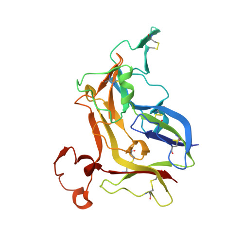
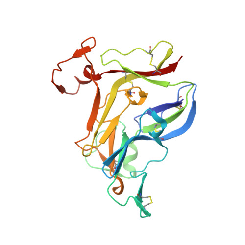
Crystal Structure of NC1 Domains. Structural Basis for Type IV Collagen Assembly in Basement Membranes

Sundaramoorthy, M.Meiyappan, M.Todd, P.Hudson, B.G.

(2002) J Biological Chem 277: 31142-31153

  • DOI: https://doi.org/10.1074/jbc.M201740200
  • Primary Citation Related Structures: 
    1M3D

  • PubMed Abstract: 

    Type IV collagen, which is present in all metazoan, exists as a family of six homologous alpha(IV) chains, alpha1-alpha6, in mammals. The six chains assemble into three different triple helical protomers and self-associate as three distinct networks. The network underlies all epithelia as a component of basement membranes, which play important roles in cell adhesion, growth, differentiation, tissue repair and molecular ultrafiltration. The specificity of both protomer and network assembly is governed by amino acid sequences of the C-terminal noncollagenous (NC1) domain of each chain. In this study, the structural basis for protomer and network assembly was investigated by determining the crystal structure of the ubiquitous [(alpha1)(2).alpha2](2) NC1 hexamer of bovine lens capsule basement membrane at 2.0 A resolution. The NC1 monomer folds into a novel tertiary structure. The (alpha1)(2).alpha2 trimer is organized through the unique three-dimensional domain swapping interactions. The differences in the primary sequences of the hypervariable region manifest in different secondary structures, which determine the chain specificity at the monomer-monomer interfaces. The trimer-trimer interface is stabilized by the extensive hydrophobic and hydrophilic interactions without a need for disulfide cross-linking.


  • Organizational Affiliation
    • Department of Biochemistry and Molecular Biology, University of Kansas Medical Center, Kansas City, KS 66160-7421, USA.

Macromolecule Content 

  • Total Structure Weight: 307.85 kDa 
  • Atom Count: 22,023 
  • Modeled Residue Count: 2,681 
  • Deposited Residue Count: 2,740 
  • Unique protein chains: 2

Macromolecules

Find similar proteins by:|  3D Structure
Entity ID: 1
MoleculeChains  Sequence LengthOrganismDetailsImage
Type IV Collagen Noncollagenous Domain- Alpha1
A, B, D, E, G
A, B, D, E, G, H, J, K
229Bos taurusMutation(s): 0 
UniProt
Find proteins for Q7SIB2 (Bos taurus)
Explore Q7SIB2 
Go to UniProtKB:  Q7SIB2
Entity Groups
Sequence Clusters30% Identity50% Identity70% Identity90% Identity95% Identity100% Identity
UniProt GroupQ7SIB2
Sequence Annotations
Expand
Reference Sequence
Find similar proteins by:|  3D Structure
Entity ID: 2
MoleculeChains  Sequence LengthOrganismDetailsImage
Type IV Collagen Noncollagenous Domain- Alpha2
C, F, I, L
227Bos taurusMutation(s): 0 
UniProt
Find proteins for Q7SIB3 (Bos taurus)
Explore Q7SIB3 
Go to UniProtKB:  Q7SIB3
Entity Groups
Sequence Clusters30% Identity50% Identity70% Identity90% Identity95% Identity100% Identity
UniProt GroupQ7SIB3
Sequence Annotations
Expand
Reference Sequence

Small Molecules

Ligands 3 Unique
IDChains Name / Formula / InChI Key2D Diagram3D Interactions
LU

Query on LU


Download:Ideal Coordinates CCD File
EB [auth L],
HA [auth F],
TA [auth I],
X [auth C]
LUTETIUM (III) ION
Lu
PSDMOPINLDTFSZ-UHFFFAOYSA-N
GOL

Query on GOL


Download:Ideal Coordinates CCD File
DA [auth D]
DB [auth K]
GA [auth E]
NA [auth G]
Q [auth A]
DA [auth D],
DB [auth K],
GA [auth E],
NA [auth G],
Q [auth A],
SA [auth H],
W [auth B],
ZA [auth J]
GLYCEROL
C3 H8 O3
PEDCQBHIVMGVHV-UHFFFAOYSA-N
BR

Query on BR


Download:Ideal Coordinates CCD File
AA [auth C]
AB [auth K]
BA [auth C]
BB [auth K]
CA [auth D]
AA [auth C],
AB [auth K],
BA [auth C],
BB [auth K],
CA [auth D],
CB [auth K],
EA [auth E],
FA [auth E],
FB [auth L],
GB [auth L],
HB [auth L],
IA [auth F],
JA [auth F],
KA [auth G],
LA [auth G],
M [auth A],
MA [auth G],
N [auth A],
O [auth A],
OA [auth H],
P [auth A],
PA [auth H],
QA [auth H],
R [auth B],
RA [auth H],
S [auth B],
T [auth B],
U [auth B],
UA [auth I],
V [auth B],
VA [auth I],
WA [auth I],
XA [auth J],
Y [auth C],
YA [auth J],
Z [auth C]
BROMIDE ION
Br
CPELXLSAUQHCOX-UHFFFAOYSA-M

Experimental Data & Validation

Experimental Data

  • Method: X-RAY DIFFRACTION
  • Resolution: 2.00 Å
  • R-Value Free:  0.197 (Depositor), 0.196 (DCC) 
  • R-Value Work:  0.169 (Depositor), 0.169 (DCC) 
  • R-Value Observed: 0.171 (Depositor) 
Space Group: P 1 21 1
Unit Cell:
Length ( Å )Angle ( ˚ )
a = 80.069α = 90
b = 137.963β = 90.31
c = 127.131γ = 90
Software Package:
Software NamePurpose
DENZOdata reduction
SCALEPACKdata scaling
SOLVEphasing
CNSrefinement

Structure Validation

View Full Validation Report


Entry History 

Deposition Data

Revision History  (Full details and data files)

  • Version 1.0: 2003-01-07
    Type: Initial release
  • Version 1.1: 2008-04-28
    Changes: Version format compliance
  • Version 1.2: 2011-07-13
    Changes: Non-polymer description, Version format compliance
  • Version 1.3: 2024-10-16
    Changes: Data collection, Database references, Derived calculations, Structure summary