1M2R | pdb_00001m2r

Crystal structure of 5,8-di-amino-1,4-di-hydroxy-anthraquinone/CK2 kinase complex


Experimental Data Snapshot

  • Method: X-RAY DIFFRACTION
  • Resolution: 1.70 Å
  • R-Value Free: 
    0.216 (Depositor) 
  • R-Value Work: 
    0.190 (Depositor) 
  • R-Value Observed: 
    0.190 (Depositor) 

wwPDB Validation 3D Report Full Report

Validation slider image for 1M2R

This is version 1.3 of the entry. See complete history

Literature

Inhibition of protein kinase CK2 by anthraquinone-related compounds. A structural insight

De Moliner, E.Sarno, S.Moro, S.Zanotti, G.Battistutta, R.Pinna, L.A.

(2003) J Biological Chem 278: 1831-1836

  • DOI: https://doi.org/10.1074/jbc.M209367200
  • Primary Citation Related Structures: 
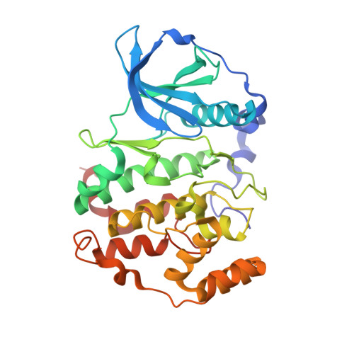
    1M2P, 1M2Q, 1M2R

  • PubMed Abstract: 

    Protein kinases play key roles in signal transduction and therefore are among the most attractive targets for drug design. The pharmacological aptitude of protein kinase inhibitors is highlighted by the observation that various diseases with special reference to cancer are because of the abnormal expression/activity of individual kinases. The resolution of the three-dimensional structure of the target kinase in complex with inhibitors is often the starting point for the rational design of this kind of drugs, some of which are already in advanced clinical trial or even in clinical practice. Here we present and discuss three new crystal structures of ATP site-directed inhibitors in complex with "casein kinase-2" (CK2), a constitutively active protein kinase implicated in a variety of cellular functions and misfunctions. With the help of theoretical calculations, we disclose some key features underlying the inhibitory efficiency of anthraquinone derivatives, outlining three different binding modes into the active site. In particular, we show that a nitro group in a hydroxyanthraquinone scaffold decreases the inhibitory constants K(i) because of electron-withdrawing and resonance effects that enhance the polarization of hydroxylic substituents in paraposition.


  • Organizational Affiliation
    • Department of Organic Chemistry, University of Padova, Padova 35131, Italy.

Macromolecule Content 

  • Total Structure Weight: 38.94 kDa 
  • Atom Count: 2,953 
  • Modeled Residue Count: 327 
  • Deposited Residue Count: 327 
  • Unique protein chains: 1

Macromolecules

Find similar proteins by:|  3D Structure
Entity ID: 1
MoleculeChains  Sequence LengthOrganismDetailsImage
CASEIN kinase II, alpha chain327Zea maysMutation(s): 0 
EC: 2.7.1.37 (PDB Primary Data), 2.7.11.1 (UniProt)
UniProt
Find proteins for P28523 (Zea mays)
Explore P28523 
Go to UniProtKB:  P28523
Entity Groups
Sequence Clusters30% Identity50% Identity70% Identity90% Identity95% Identity100% Identity
UniProt GroupP28523
Sequence Annotations
Expand
Reference Sequence

Small Molecules

Ligands 1 Unique
IDChains Name / Formula / InChI Key2D Diagram3D Interactions
MNY

Query on MNY


Download:Ideal Coordinates CCD File
B [auth A]5,8-DI-AMINO-1,4-DIHYDROXY-ANTHRAQUINONE
C14 H10 N2 O4
QVEMRPAUHFWHRV-UHFFFAOYSA-N
Binding Affinity Annotations 
IDSourceBinding Affinity
MNY BindingDB:  1M2R Ki: 350 (nM) from 1 assay(s)

Experimental Data & Validation

Experimental Data

  • Method: X-RAY DIFFRACTION
  • Resolution: 1.70 Å
  • R-Value Free:  0.216 (Depositor) 
  • R-Value Work:  0.190 (Depositor) 
  • R-Value Observed: 0.190 (Depositor) 
Space Group: C 1 2 1
Unit Cell:
Length ( Å )Angle ( ˚ )
a = 143.11α = 90
b = 51.83β = 99.65
c = 44.71γ = 90
Software Package:
Software NamePurpose
MOSFLMdata reduction
SCALAdata scaling
AMoREphasing
CNSrefinement
CCP4data scaling

Structure Validation

View Full Validation Report


Entry History 

Deposition Data

Revision History  (Full details and data files)

  • Version 1.0: 2003-06-17
    Type: Initial release
  • Version 1.1: 2008-04-28
    Changes: Version format compliance
  • Version 1.2: 2011-07-13
    Changes: Version format compliance
  • Version 1.3: 2024-02-14
    Changes: Data collection, Database references, Derived calculations