1LZ1 | pdb_00001lz1

REFINEMENT OF HUMAN LYSOZYME AT 1.5 ANGSTROMS RESOLUTION. ANALYSIS OF NON-BONDED AND HYDROGEN-BOND INTERACTIONS


Experimental Data Snapshot

  • Method: X-RAY DIFFRACTION
  • Resolution: 1.50 Å
  • R-Value Work: 
    0.177 (Depositor) 

wwPDB Validation 3D Report Full Report

Validation slider image for 1LZ1

This is version 1.4 of the entry. See complete history

Literature

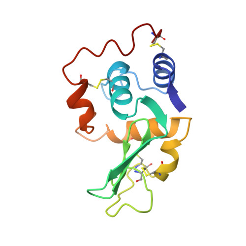
Refinement of human lysozyme at 1.5 A resolution analysis of non-bonded and hydrogen-bond interactions.

Artymiuk, P.J.Blake, C.C.

(1981) J Mol Biology 152: 737-762

Macromolecule Content 

  • Total Structure Weight: 14.72 kDa 
  • Atom Count: 1,029 
  • Modeled Residue Count: 130 
  • Deposited Residue Count: 130 
  • Unique protein chains: 1

Macromolecules

Find similar proteins by:|  3D Structure
Entity ID: 1
MoleculeChains  Sequence LengthOrganismDetailsImage
HUMAN LYSOZYME130Homo sapiensMutation(s): 0 
EC: 3.2.1.17
UniProt & NIH Common Fund Data Resources
Find proteins for P61626 (Homo sapiens)
Explore P61626 
Go to UniProtKB:  P61626
PHAROS:  P61626
GTEx:  ENSG00000090382 
Entity Groups
Sequence Clusters30% Identity50% Identity70% Identity90% Identity95% Identity100% Identity
UniProt GroupP61626
Sequence Annotations
Expand
Reference Sequence

Experimental Data & Validation

Experimental Data

  • Method: X-RAY DIFFRACTION
  • Resolution: 1.50 Å
  • R-Value Work:  0.177 (Depositor) 
Space Group: P 21 21 21
Unit Cell:
Length ( Å )Angle ( ˚ )
a = 57.13α = 90
b = 60.99β = 90
c = 32.89γ = 90

Structure Validation

View Full Validation Report


Entry History 

Deposition Data

Revision History  (Full details and data files)

  • Version 1.0: 1985-01-02
    Type: Initial release
  • Version 1.1: 2008-03-03
    Changes: Version format compliance
  • Version 1.2: 2011-07-13
    Changes: Version format compliance
  • Version 1.3: 2017-11-29
    Changes: Derived calculations, Other
  • Version 1.4: 2024-10-30
    Changes: Data collection, Database references, Structure summary