1LY2 | pdb_00001ly2

Crystal structure of unliganded human CD21 SCR1-SCR2 (Complement receptor type 2)


Experimental Data Snapshot

  • Method: X-RAY DIFFRACTION
  • Resolution: 1.80 Å
  • R-Value Free: 
    0.234 (Depositor), 0.231 (DCC) 
  • R-Value Work: 
    0.179 (Depositor), 0.177 (DCC) 

Starting Model: experimental
View more details

wwPDB Validation 3D Report Full Report

Validation slider image for 1LY2

Ligand Structure Quality Assessment 


This is version 1.5 of the entry. See complete history

Literature

The crystal structure of human CD21: Implications for Epstein-Barr virus and C3d binding.

Prota, A.E.Sage, D.R.Stehle, T.Fingeroth, J.D.

(2002) Proc Natl Acad Sci U S A 99: 10641-10646

  • DOI: https://doi.org/10.1073/pnas.162360499
  • Primary Citation Related Structures: 
    1LY2

  • PubMed Abstract: 

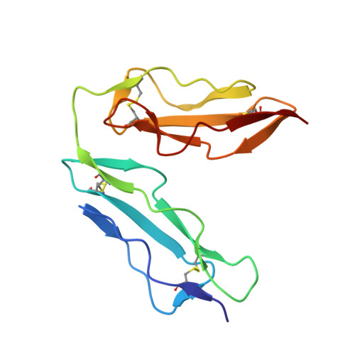
    Human complement receptor type 2 (CD21) is the cellular receptor for Epstein-Barr virus (EBV), a human tumor virus. The N-terminal two short consensus repeats (SCR1-SCR2) of the receptor interact with the EBV glycoprotein gp350/220 and also with the natural CD21 ligand C3d. Here we present the crystal structure of the CD21 SCR1-SCR2 fragment in the absence of ligand and demonstrate that it is able to bind EBV. Based on a functional analysis of wild-type and mutant CD21 and molecular modeling, we identify a likely region for EBV attachment and demonstrate that this region is not involved in the interaction with C3d. A comparison with the previously determined structure of CD21 SCR1-SCR2 in complex with C3d shows that, in both cases, CD21 assumes compact V-shaped conformations. However, our analysis reveals a surprising degree of flexibility at the SCR1-SCR2 interface, suggesting interactions between the two domains are not specific. We present evidence that the V-shaped conformation is induced by deglycosylation of the protein, and that physiologic glycosylation of CD21 would result in a more extended conformation, perhaps with additional epitopes for C3d binding.


  • Organizational Affiliation
    • Harvard Medical School, Division of Experimental Medicine and Infectious Diseases, Beth Israel Deaconess Medical Center, Harvard Institutes of Medicine, 4 Blackfan Circle, Boston, MA 02115, USA.

Macromolecule Content 

  • Total Structure Weight: 14.7 kDa 
  • Atom Count: 1,170 
  • Modeled Residue Count: 130 
  • Deposited Residue Count: 130 
  • Unique protein chains: 1

Macromolecules

Find similar proteins by:|  3D Structure
Entity ID: 1
MoleculeChains  Sequence LengthOrganismDetailsImage
complement receptor type 2130Homo sapiensMutation(s): 0 
Gene Names: CR2
UniProt & NIH Common Fund Data Resources
Find proteins for P20023 (Homo sapiens)
Explore P20023 
Go to UniProtKB:  P20023
PHAROS:  P20023
GTEx:  ENSG00000117322 
Entity Groups
Sequence Clusters30% Identity50% Identity70% Identity90% Identity95% Identity100% Identity
UniProt GroupP20023
Glycosylation
Glycosylation Sites: 2Go to GlyGen: P20023-1
Sequence Annotations
Expand
Reference Sequence

Experimental Data & Validation

Experimental Data

  • Method: X-RAY DIFFRACTION
  • Resolution: 1.80 Å
  • R-Value Free:  0.234 (Depositor), 0.231 (DCC) 
  • R-Value Work:  0.179 (Depositor), 0.177 (DCC) 
Space Group: P 1
Unit Cell:
Length ( Å )Angle ( ˚ )
a = 31.6α = 91.51
b = 33.27β = 111.44
c = 42.11γ = 117.81
Software Package:
Software NamePurpose
DENZOdata reduction
SCALEPACKdata scaling
AMoREphasing
X-PLORrefinement

Structure Validation

View Full Validation Report


Ligand Structure Quality Assessment 


Entry History 

Deposition Data

Revision History  (Full details and data files)

  • Version 1.0: 2002-07-05
    Type: Initial release
  • Version 1.1: 2008-04-28
    Changes: Version format compliance
  • Version 1.2: 2011-07-13
    Changes: Non-polymer description, Version format compliance
  • Version 1.3: 2020-07-29
    Type: Remediation
    Reason: Carbohydrate remediation
    Changes: Data collection, Database references, Derived calculations, Structure summary
  • Version 1.4: 2024-04-03
    Changes: Data collection, Database references, Refinement description, Structure summary
  • Version 1.5: 2024-11-20
    Changes: Structure summary