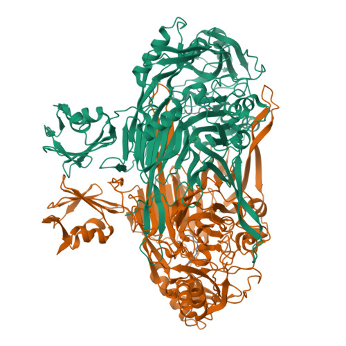
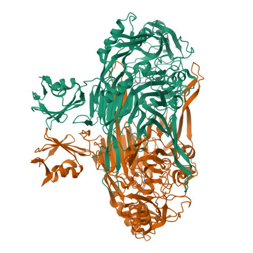
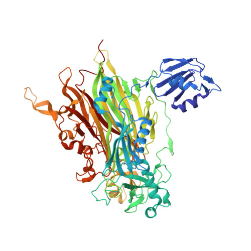
1LVN

CRYSTAL STRUCTURE OF E. COLI AMINE OXIDASE COMPLEXED WITH TRANYLCYPROMINE


Experimental Data Snapshot

  • Method: X-RAY DIFFRACTION
  • Resolution: 2.40 Å
  • R-Value Free: 0.229 
  • R-Value Work: 0.185 
  • R-Value Observed: 0.185 

Starting Model: experimental
View more details

wwPDB Validation   3D Report Full Report


This is version 2.1 of the entry. See complete history


Literature

Medical implications from the crystal structure of a copper-containing amine oxidase complexed with the antidepressant drug tranylcypromine.

Wilmot, C.M.Saysell, C.G.Blessington, A.Conn, D.A.Kurtis, C.R.McPherson, M.J.Knowles, P.F.Phillips, S.E.

(2004) FEBS Lett 576: 301-305

  • DOI: https://doi.org/10.1016/j.febslet.2004.09.031
  • Primary Citation of Related Structures:  
    1LVN

  • PubMed Abstract: 

    The X-ray crystal structure of the copper-containing quinoprotein amine oxidase from E. coli has been determined in complex with the antidepressant drug tranylcypromine to 2.4 A resolution. The drug is a racemic mix of two enantiomers, but only one is seen bound to the enzyme. The other enantiomer is not acting as a substrate for the enzyme as no catalytic activity was detected when the enzyme was initially exposed to the drug. The inhibition of human copper amine oxidases could be a source of side-effects in its use as an antidepressant to inhibit the flavin-containing monoamine oxidases in the brain.


  • Organizational Affiliation

    Astbury Centre for Structural Molecular Biology, School of Biochemistry and Molecular Biology, University of Leeds, Leeds LS2 9JT, UK. wilmo004@umn.edu


Macromolecules
Find similar proteins by:  (by identity cutoff)  |  3D Structure
Entity ID: 1
MoleculeChains Sequence LengthOrganismDetailsImage
Copper amine oxidase
A, B
727Escherichia coliMutation(s): 1 
EC: 1.4.3.6 (PDB Primary Data), 1.4.3.21 (UniProt)
UniProt
Find proteins for P46883 (Escherichia coli (strain K12))
Explore P46883 
Go to UniProtKB:  P46883
Entity Groups  
Sequence Clusters30% Identity50% Identity70% Identity90% Identity95% Identity100% Identity
UniProt GroupP46883
Sequence Annotations
Expand
  • Reference Sequence
Experimental Data & Validation

Experimental Data

  • Method: X-RAY DIFFRACTION
  • Resolution: 2.40 Å
  • R-Value Free: 0.229 
  • R-Value Work: 0.185 
  • R-Value Observed: 0.185 
  • Space Group: P 21 21 21
Unit Cell:
Length ( Å )Angle ( ˚ )
a = 135.236α = 90
b = 166.482β = 90
c = 79.628γ = 90
Software Package:
Software NamePurpose
MOSFLMdata reduction
ROTAVATAdata reduction
CNSrefinement
CCP4data scaling
ROTAVATAdata scaling
CNSphasing

Structure Validation

View Full Validation Report


Entry History 

Deposition Data

Revision History  (Full details and data files)

  • Version 1.0: 2003-08-05
    Type: Initial release
  • Version 1.1: 2008-04-28
    Changes: Version format compliance
  • Version 1.2: 2011-07-13
    Changes: Version format compliance
  • Version 1.3: 2017-10-11
    Changes: Refinement description
  • Version 2.0: 2023-11-15
    Changes: Atomic model, Data collection, Database references, Derived calculations, Refinement description
  • Version 2.1: 2024-11-13
    Changes: Structure summary