1LTQ | pdb_00001ltq

CRYSTAL STRUCTURE OF T4 POLYNUCLEOTIDE KINASE


Experimental Data Snapshot

  • Method: X-RAY DIFFRACTION
  • Resolution: 2.33 Å
  • R-Value Free: 
    0.261 (Depositor) 
  • R-Value Work: 
    0.236 (Depositor) 
  • R-Value Observed: 
    0.238 (Depositor) 

wwPDB Validation 3D Report Full Report

Validation slider image for 1LTQ

This is version 1.3 of the entry. See complete history

Literature

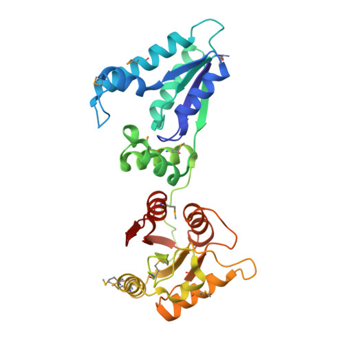
Structure of a tRNA repair enzyme and molecular biology workhorse: T4 polynucleotide kinase.

Galburt, E.A.Pelletier, J.Wilson, G.Stoddard, B.L.

(2002) Structure 10: 1249-1260

  • DOI: https://doi.org/10.1016/s0969-2126(02)00835-3
  • Primary Citation Related Structures: 
    1LTQ

  • PubMed Abstract: 

    T4 phage polynucleotide kinase (PNK) was identified over 35 years ago and has become a staple reagent for molecular biologists. The enzyme displays 5'-hydroxyl kinase, 3'-phosphatase, and 2',3'-cyclic phosphodiesterase activities against a wide range of substrates. These activities modify the ends of nicked tRNA generated by a bacterial response to infection and facilitate repair by T4 RNA ligase. DNA repair enzymes that share conserved motifs with PNK have been identified in eukaryotes. PNK contains two functionally distinct structural domains and forms a homotetramer. The C-terminal phosphatase domain is homologous to the L-2-haloacid dehalogenase family and the N-terminal kinase domain is homologous to adenylate kinase. The active sites have been characterized through structural homology analyses and visualization of bound substrate.


  • Organizational Affiliation
    • Fred Hutchinson Cancer Research Center and The Graduate Program in Biomolecular Structure and Design, University of Washington, 1100 Fairview Avenue North, A3-023, Seattle, WA 98109, USA.

Macromolecule Content 

  • Total Structure Weight: 35.74 kDa 
  • Atom Count: 2,532 
  • Modeled Residue Count: 298 
  • Deposited Residue Count: 301 
  • Unique protein chains: 1

Macromolecules

Find similar proteins by:|  3D Structure
Entity ID: 1
MoleculeChains  Sequence LengthOrganismDetailsImage
POLYNUCLEOTIDE KINASE301Tequatrovirus T4Mutation(s): 12 
EC: 2.7.1.78 (PDB Primary Data), 3.1.3.34 (UniProt)
UniProt
Find proteins for P06855 (Enterobacteria phage T4)
Explore P06855 
Go to UniProtKB:  P06855
Entity Groups
Sequence Clusters30% Identity50% Identity70% Identity90% Identity95% Identity100% Identity
UniProt GroupP06855
Sequence Annotations
Expand
Reference Sequence

Small Molecules

Ligands 2 Unique
IDChains Name / Formula / InChI Key2D Diagram3D Interactions
ADP

Query on ADP


Download:Ideal Coordinates CCD File
B [auth A]ADENOSINE-5'-DIPHOSPHATE
C10 H15 N5 O10 P2
XTWYTFMLZFPYCI-KQYNXXCUSA-N
DMS

Query on DMS


Download:Ideal Coordinates CCD File
C [auth A]DIMETHYL SULFOXIDE
C2 H6 O S
IAZDPXIOMUYVGZ-UHFFFAOYSA-N
Modified Residues  1 Unique
IDChains TypeFormula2D DiagramParent
MSE
Query on MSE
A
L-PEPTIDE LINKINGC5 H11 N O2 SeMET

Experimental Data & Validation

Experimental Data

  • Method: X-RAY DIFFRACTION
  • Resolution: 2.33 Å
  • R-Value Free:  0.261 (Depositor) 
  • R-Value Work:  0.236 (Depositor) 
  • R-Value Observed: 0.238 (Depositor) 
Space Group: I 2 2 2
Unit Cell:
Length ( Å )Angle ( ˚ )
a = 78.585α = 90
b = 93.684β = 90
c = 124.165γ = 90
Software Package:
Software NamePurpose
HKL-2000data collection
SCALEPACKdata scaling
CNSrefinement
HKL-2000data reduction
CNSphasing

Structure Validation

View Full Validation Report


Entry History 

Deposition Data

Revision History  (Full details and data files)

  • Version 1.0: 2002-10-09
    Type: Initial release
  • Version 1.1: 2008-04-28
    Changes: Version format compliance
  • Version 1.2: 2011-07-13
    Changes: Derived calculations, Version format compliance
  • Version 1.3: 2024-10-09
    Changes: Data collection, Database references, Derived calculations, Structure summary