1LTO | pdb_00001lto

Human alpha1-tryptase


Experimental Data Snapshot

  • Method: X-RAY DIFFRACTION
  • Resolution: 2.20 Å
  • R-Value Free: 
    0.267 (Depositor) 
  • R-Value Work: 
    0.214 (Depositor) 
  • R-Value Observed: 
    0.214 (Depositor) 

Starting Model: experimental
View more details

wwPDB Validation 3D Report Full Report

Validation slider image for 1LTO

This is version 1.4 of the entry. See complete history

Literature

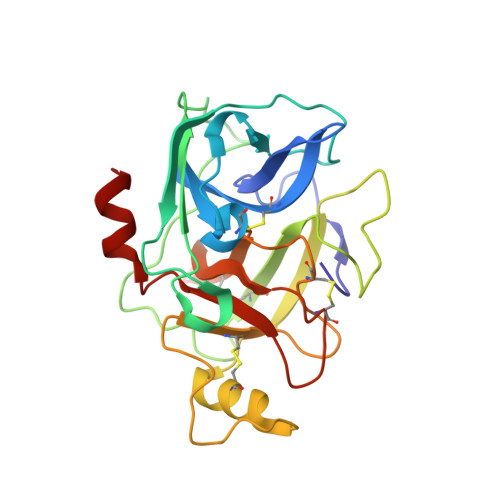
The Crystal Structure of Human alpha1-Tryptase Reveals a Blocked Substrate-binding Region

Marquardt, U.Zettl, F.Huber, R.Bode, W.Sommerhoff, C.P.

(2002) J Mol Biology 321: 491-502

  • DOI: https://doi.org/10.1016/s0022-2836(02)00625-3
  • Primary Citation Related Structures: 
    1LTO

  • PubMed Abstract: 

    Human mast cell tryptases represent a subfamily of trypsin-like serine proteinases implicated in asthma. Unlike beta-tryptases, alpha-tryptases apparently are proteolytically inactive. We have solved the 2.2A crystal structure of mature human alpha1-tryptase. It reveals a frame-like tetrameric architecture that, surprisingly, does not require heparin-binding for stability. In marked contrast to beta2-tryptase, the Ser214-Gly219 segment, which normally provides the template for substrate binding, is kinked in alpha-tryptase, thereby blocking its non-primed subsites. This so far unobserved subsite distortion is incompatible with productive substrate binding and processing. alpha-Tryptase apparently is trapped in this off-conformation by repulsions and attractions of the Asp216 side-chain. However, proteolytic activity could be generated by an induced-fit mechanism.


  • Organizational Affiliation
    • Max-Planck-Institut für Biochemie, Abteilung Strukturforschung, Am Klopferspitz 18a, D-82152 Martinsried bei, München, Germany.

Macromolecule Content 

  • Total Structure Weight: 110.93 kDa 
  • Atom Count: 8,343 
  • Modeled Residue Count: 972 
  • Deposited Residue Count: 980 
  • Unique protein chains: 1

Macromolecules

Find similar proteins by:|  3D Structure
Entity ID: 1
MoleculeChains  Sequence LengthOrganismDetailsImage
alpha tryptase I
A, B, C, D
245Homo sapiensMutation(s): 0 
EC: 3.4.21.59
UniProt & NIH Common Fund Data Resources
Find proteins for Q15661 (Homo sapiens)
Explore Q15661 
Go to UniProtKB:  Q15661
PHAROS:  Q15661
GTEx:  ENSG00000172236 
Entity Groups
Sequence Clusters30% Identity50% Identity70% Identity90% Identity95% Identity100% Identity
UniProt GroupQ15661
Sequence Annotations
Expand
Reference Sequence

Experimental Data & Validation

Experimental Data

  • Method: X-RAY DIFFRACTION
  • Resolution: 2.20 Å
  • R-Value Free:  0.267 (Depositor) 
  • R-Value Work:  0.214 (Depositor) 
  • R-Value Observed: 0.214 (Depositor) 
Space Group: P 21 21 21
Unit Cell:
Length ( Å )Angle ( ˚ )
a = 91.441α = 90
b = 110.685β = 90
c = 130.092γ = 90
Software Package:
Software NamePurpose
MOSFLMdata reduction
SCALAdata scaling
AMoREphasing
CNSrefinement
CCP4data scaling

Structure Validation

View Full Validation Report


Entry History 

Deposition Data

Revision History  (Full details and data files)

  • Version 1.0: 2003-05-20
    Type: Initial release
  • Version 1.1: 2008-04-28
    Changes: Version format compliance
  • Version 1.2: 2011-07-13
    Changes: Version format compliance
  • Version 1.3: 2024-04-03
    Changes: Data collection, Database references, Refinement description
  • Version 1.4: 2024-10-09
    Changes: Structure summary