1LQX

Crystal structure of V45E mutant of cytochrome b5


Experimental Data Snapshot

  • Method: X-RAY DIFFRACTION
  • Resolution: 1.80 Å
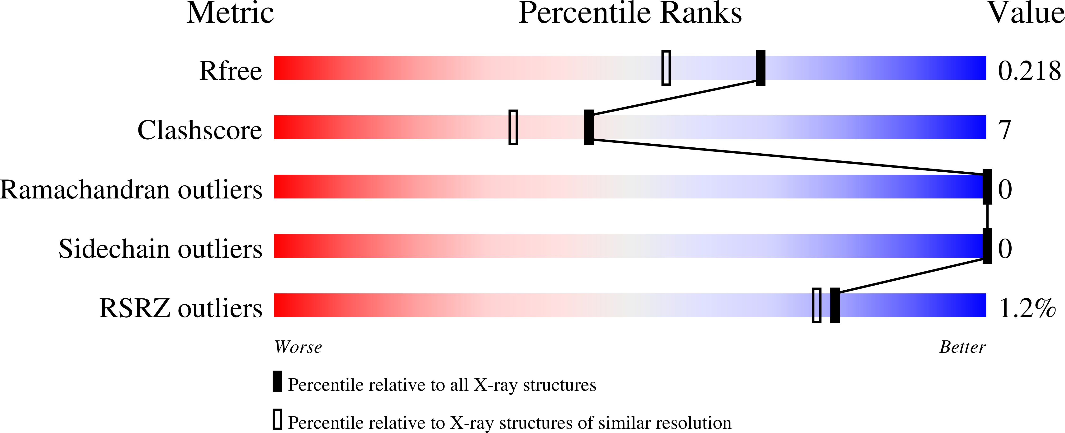
  • R-Value Free: 0.222 
  • R-Value Work: 0.189 
  • R-Value Observed: 0.189 

Starting Model: experimental
View more details

wwPDB Validation   3D Report Full Report


Ligand Structure Quality Assessment 


This is version 1.4 of the entry. See complete history


Literature

Structures of V45E and V45Y mutants and structure comparison of a variety of cytochrome b5 mutants.

Gan, J.H.Wu, J.Wang, Z.Q.Wang, Y.H.Huang, Z.X.Xia, Z.X.

(2002) Acta Crystallogr D Biol Crystallogr 58: 1298-1306

  • DOI: https://doi.org/10.1107/s0907444902010016
  • Primary Citation of Related Structures:  
    1LQX, 1LR6

  • PubMed Abstract: 

    Val45 is a highly conserved residue and a component of the heme-pocket wall of cytochrome b(5). The crystal structures of cytochrome b(5) mutants V45E and V45Y have been determined at high resolution. Their overall structures were very similar to that of the wild-type protein. However, Val45 of the wild-type protein points towards the heme, but the large side chains of both Glu45 and Tyr45 of the mutants point towards the solvent. A channel is thus opened and the hydrophobicity of the heme pocket is decreased. The rotation of the porphyrin ring and the conformational change of the axial ligand His39 in the V45Y mutant indicate that the microenvironment of the heme is disturbed because of the mutation. The binding constants and the electron-transfer rates between cytochrome b(5) and cytochrome c decrease owing to the mutation, which can be accounted for by molecular modeling: the inter-iron distances increase in order to eliminate the unreasonably close contacts resulting from the large volumes of the mutated side chains. The influence of the mutations on the redox potentials and protein stability is also discussed. The structures of seven mutants of cytochrome b(5) are compared with each other and the effects of these mutations on the protein properties and functions are summarized.


  • Organizational Affiliation

    State Key Laboratory of Bioorganic and Natural Products Chemistry, Shanghai Institute of Organic Chemistry, Chinese Academy of Sciences, Shanghai 200032, People's Republic of China.


Macromolecules
Find similar proteins by:  (by identity cutoff)  |  3D Structure
Entity ID: 1
MoleculeChains Sequence LengthOrganismDetailsImage
cytochrome b582Bos taurusMutation(s): 1 
UniProt
Find proteins for P00171 (Bos taurus)
Explore P00171 
Go to UniProtKB:  P00171
Entity Groups  
Sequence Clusters30% Identity50% Identity70% Identity90% Identity95% Identity100% Identity
UniProt GroupP00171
Sequence Annotations
Expand
  • Reference Sequence
Small Molecules
Ligands 1 Unique
IDChains Name / Formula / InChI Key2D Diagram3D Interactions
HEM
Query on HEM

Download Ideal Coordinates CCD File 
B [auth A]PROTOPORPHYRIN IX CONTAINING FE
C34 H32 Fe N4 O4
KABFMIBPWCXCRK-RGGAHWMASA-L
Experimental Data & Validation

Experimental Data

  • Method: X-RAY DIFFRACTION
  • Resolution: 1.80 Å
  • R-Value Free: 0.222 
  • R-Value Work: 0.189 
  • R-Value Observed: 0.189 
  • Space Group: C 1 2 1
Unit Cell:
Length ( Å )Angle ( ˚ )
a = 70.913α = 90
b = 40.338β = 111.57
c = 39.153γ = 90
Software Package:
Software NamePurpose
CNSrefinement
DENZOdata reduction
SCALEPACKdata scaling
CNSphasing

Structure Validation

View Full Validation Report


Ligand Structure Quality Assessment 


Entry History 

Deposition Data

Revision History  (Full details and data files)

  • Version 1.0: 2002-09-04
    Type: Initial release
  • Version 1.1: 2008-04-28
    Changes: Version format compliance
  • Version 1.2: 2011-07-13
    Changes: Version format compliance
  • Version 1.3: 2021-11-10
    Changes: Database references, Derived calculations
  • Version 1.4: 2023-10-25
    Changes: Data collection, Refinement description