1LPN | pdb_00001lpn

ANALOGS OF REACTION INTERMEDIATES IDENTIFY A UNIQUE SUBSTRATE BINDING SITE IN CANDIDA RUGOSA LIPASE


Experimental Data Snapshot

  • Method: X-RAY DIFFRACTION
  • Resolution: 2.18 Å
  • R-Value Work: 
    0.151 (Depositor), 0.161 (DCC) 
  • R-Value Observed: 
    0.151 (Depositor) 

wwPDB Validation 3D Report Full Report

Validation slider image for 1LPN

Ligand Structure Quality Assessment 


This is version 2.1 of the entry. See complete history

Literature

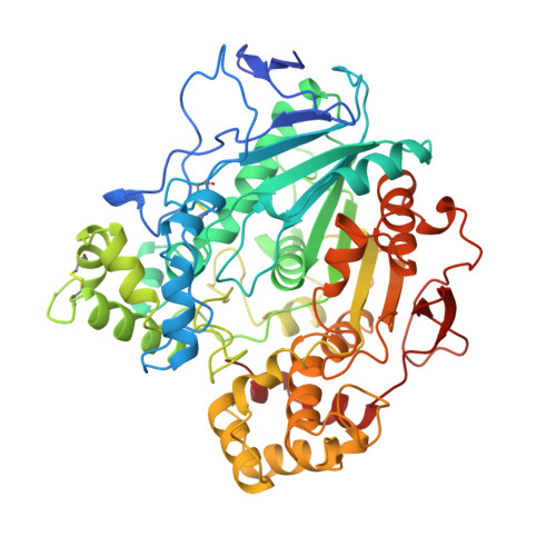
Analogs of reaction intermediates identify a unique substrate binding site in Candida rugosa lipase.

Grochulski, P.Bouthillier, F.Kazlauskas, R.J.Serreqi, A.N.Schrag, J.D.Ziomek, E.Cygler, M.

(1994) Biochemistry 33: 3494-3500

  • DOI: https://doi.org/10.1021/bi00178a005
  • Primary Citation Related Structures: 
    1LPN, 1LPO, 1LPP

  • PubMed Abstract: 

    The structures of Candida rugosa lipase-inhibitor complexes demonstrate that the scissile fatty acyl chain is bound in a narrow, hydrophobic tunnel which is unique among lipases studied to date. Modeling of triglyceride binding suggests that the bound lipid must adopt a "tuning fork" conformation. The complexes, analogs of tetrahedral intermediates of the acylation and deacylation steps of the reaction pathway, localize the components of the oxyanion hole and define the stereochemistry of ester hydrolysis. Comparison with other lipases suggests that the positioning of the scissile fatty acyl chain and ester bond and the stereochemistry of hydrolysis are the same in all lipases which share the alpha/beta-hydrolase fold.


  • Organizational Affiliation
    • Biotechnology Research Institute, National Research Council of Canada, Montréal, Quebec.

Macromolecule Content 

  • Total Structure Weight: 59.81 kDa 
  • Atom Count: 4,211 
  • Modeled Residue Count: 534 
  • Deposited Residue Count: 549 
  • Unique protein chains: 1

Macromolecules

Find similar proteins by:|  3D Structure
Entity ID: 1
MoleculeChains  Sequence LengthOrganismDetailsImage
LIPASE549Diutina rugosaMutation(s): 0 
EC: 3.1.1.3
UniProt
Find proteins for P20261 (Diutina rugosa)
Explore P20261 
Go to UniProtKB:  P20261
Entity Groups
Sequence Clusters30% Identity50% Identity70% Identity90% Identity95% Identity100% Identity
UniProt GroupP20261
Glycosylation
Glycosylation Sites: 2
Sequence Annotations
Expand
Reference Sequence

Oligosaccharides

Help  
Entity ID: 2
MoleculeChains Length2D Diagram GlycosylationD Interactions
2-acetamido-2-deoxy-beta-D-glucopyranose-(1-4)-2-acetamido-2-deoxy-beta-D-glucopyranose
B
2N-Glycosylation
Glycosylation Resources
GlyTouCan: G42666HT
GlyCosmos: G42666HT
GlyGen: G42666HT

Experimental Data & Validation

Experimental Data

  • Method: X-RAY DIFFRACTION
  • Resolution: 2.18 Å
  • R-Value Work:  0.151 (Depositor), 0.161 (DCC) 
  • R-Value Observed: 0.151 (Depositor) 
Space Group: C 2 2 21
Unit Cell:
Length ( Å )Angle ( ˚ )
a = 65α = 90
b = 97.4β = 90
c = 176.4γ = 90
Software Package:
Software NamePurpose
X-PLORmodel building
X-PLORrefinement
X-PLORphasing

Structure Validation

View Full Validation Report


Ligand Structure Quality Assessment 


Entry History 

Deposition Data

Revision History  (Full details and data files)

  • Version 1.0: 1995-04-20
    Type: Initial release
  • Version 1.1: 2008-03-24
    Changes: Version format compliance
  • Version 1.2: 2011-07-13
    Changes: Derived calculations, Version format compliance
  • Version 2.0: 2020-07-29
    Type: Remediation
    Reason: Carbohydrate remediation
    Changes: Atomic model, Data collection, Derived calculations, Other, Structure summary
  • Version 2.1: 2024-10-30
    Changes: Data collection, Database references, Structure summary