1LOJ | pdb_00001loj

Crystal structure of a Methanobacterial Sm-like archaeal protein (SmAP1) bound to uridine-5'-monophosphate (UMP)


Experimental Data Snapshot

  • Method: X-RAY DIFFRACTION
  • Resolution: 1.90 Å
  • R-Value Free: 
    0.250 (Depositor), 0.249 (DCC) 
  • R-Value Work: 
    0.207 (Depositor), 0.207 (DCC) 
  • R-Value Observed: 
    0.207 (Depositor) 

Starting Model: experimental
View more details

wwPDB Validation 3D Report Full Report

Validation slider image for 1LOJ

Ligand Structure Quality Assessment 


This is version 2.0 of the entry. See complete history

Literature

The oligomerization and ligand-binding properties of Sm-like archaeal proteins (SmAPs)

Mura, C.Kozhukhovsky, A.Gingery, M.Phillips, M.Eisenberg, D.

(2003) Protein Sci 12: 832-847

  • DOI: https://doi.org/10.1110/ps.0224703
  • Primary Citation Related Structures: 
    1JBM, 1JRI, 1LNX, 1LOJ

  • PubMed Abstract: 

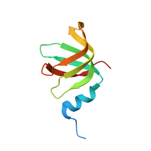
    Intron splicing is a prime example of the many types of RNA processing catalyzed by small nuclear ribonucleoprotein (snRNP) complexes. Sm proteins form the cores of most snRNPs, and thus to learn principles of snRNP assembly we characterized the oligomerization and ligand-binding properties of Sm-like archaeal proteins (SmAPs) from Pyrobaculum aerophilum (Pae) and Methanobacterium thermautotrophicum (Mth). Ultracentrifugation shows that Mth SmAP1 is exclusively heptameric in solution, whereas Pae SmAP1 forms either disulfide-bonded 14-mers or sub-heptameric states (depending on the redox potential). By electron microscopy, we show that Pae and Mth SmAP1 polymerize into bundles of well ordered fibers that probably form by head-to-tail stacking of heptamers. The crystallographic results reported here corroborate these findings by showing heptamers and 14-mers of both Mth and Pae SmAP1 in four new crystal forms. The 1.9 A-resolution structure of Mth SmAP1 bound to uridine-5'-monophosphate (UMP) reveals conserved ligand-binding sites. The likely RNA binding site in Mth agrees with that determined for Archaeoglobus fulgidus (Afu) SmAP1. Finally, we found that both Pae and Mth SmAP1 gel-shift negatively supercoiled DNA. These results distinguish SmAPs from eukaryotic Sm proteins and suggest that SmAPs have a generic single-stranded nucleic acid-binding activity.


  • Organizational Affiliation
    • Howard Hughes Medical Institute, Molecular Biology Institute, Los Angeles, California 90095-1570, USA.

Macromolecule Content 

  • Total Structure Weight: 140.58 kDa 
  • Atom Count: 8,974 
  • Modeled Residue Count: 1,047 
  • Deposited Residue Count: 1,218 
  • Unique protein chains: 1

Macromolecules

Find similar proteins by:|  3D Structure
Entity ID: 1
MoleculeChains  Sequence LengthOrganismDetailsImage
small nuclear ribonucleoprotein homolog (Sm-like)
A, B, C, D, E
A, B, C, D, E, F, G, H, I, J, K, L, M, N
87Methanothermobacter thermautotrophicus str. Delta HMutation(s): 0 
Gene Names: Mth649
UniProt
Find proteins for O26745 (Methanothermobacter thermautotrophicus (strain ATCC 29096 / DSM 1053 / JCM 10044 / NBRC 100330 / Delta H))
Explore O26745 
Go to UniProtKB:  O26745
Entity Groups
Sequence Clusters30% Identity50% Identity70% Identity90% Identity95% Identity100% Identity
UniProt GroupO26745
Sequence Annotations
Expand
Reference Sequence

Small Molecules

Ligands 4 Unique
IDChains Name / Formula / InChI Key2D Diagram3D Interactions
U5P

Query on U5P


Download:Ideal Coordinates CCD File
OA [auth N]URIDINE-5'-MONOPHOSPHATE
C9 H13 N2 O9 P
DJJCXFVJDGTHFX-XVFCMESISA-N
U

Query on U


Download:Ideal Coordinates CCD File
CA [auth H],
EA [auth I],
IA [auth K],
O [auth A],
U [auth D]
URIDINE-5'-MONOPHOSPHATE
C9 H13 N2 O9 P
DJJCXFVJDGTHFX-XVFCMESISA-N
URI

Query on URI


Download:Ideal Coordinates CCD File
AA [auth G]
GA [auth J]
KA [auth L]
MA [auth M]
Q [auth B]
AA [auth G],
GA [auth J],
KA [auth L],
MA [auth M],
Q [auth B],
S [auth C],
W [auth E],
Y [auth F]
URIDINE
C9 H12 N2 O6
DRTQHJPVMGBUCF-XVFCMESISA-N
MPD

Query on MPD


Download:Ideal Coordinates CCD File
BA [auth G]
DA [auth H]
FA [auth I]
HA [auth J]
JA [auth K]
BA [auth G],
DA [auth H],
FA [auth I],
HA [auth J],
JA [auth K],
LA [auth L],
NA [auth M],
P [auth A],
PA [auth N],
R [auth B],
T [auth C],
V [auth D],
X [auth E],
Z [auth F]
(4S)-2-METHYL-2,4-PENTANEDIOL
C6 H14 O2
SVTBMSDMJJWYQN-YFKPBYRVSA-N

Experimental Data & Validation

Experimental Data

  • Method: X-RAY DIFFRACTION
  • Resolution: 1.90 Å
  • R-Value Free:  0.250 (Depositor), 0.249 (DCC) 
  • R-Value Work:  0.207 (Depositor), 0.207 (DCC) 
  • R-Value Observed: 0.207 (Depositor) 
Space Group: P 1 21 1
Unit Cell:
Length ( Å )Angle ( ˚ )
a = 65.254α = 90
b = 109.956β = 95.81
c = 83.756γ = 90
Software Package:
Software NamePurpose
DENZOdata reduction
SCALEPACKdata scaling
EPMRphasing
CNSrefinement

Structure Validation

View Full Validation Report


Ligand Structure Quality Assessment 


Entry History 

Deposition Data

Revision History  (Full details and data files)

  • Version 1.0: 2003-03-25
    Type: Initial release
  • Version 1.1: 2008-04-28
    Changes: Version format compliance
  • Version 1.2: 2011-07-13
    Changes: Version format compliance
  • Version 1.3: 2023-08-16
    Changes: Data collection, Database references, Derived calculations, Refinement description
  • Version 2.0: 2024-10-16
    Changes: Advisory, Atomic model, Data collection, Derived calculations, Non-polymer description, Structure summary