1LLU | pdb_00001llu

THE TERNARY COMPLEX OF PSEUDOMONAS AERUGINOSA ALCOHOL DEHYDROGENASE WITH ITS COENZYME AND WEAK SUBSTRATE


Experimental Data Snapshot

  • Method: X-RAY DIFFRACTION
  • Resolution: 2.30 Å
  • R-Value Free: 
    0.246 (Depositor), 0.249 (DCC) 
  • R-Value Work: 
    0.229 (Depositor), 0.234 (DCC) 

Starting Model: experimental
View more details

wwPDB Validation 3D Report Full Report

Validation slider image for 1LLU

Ligand Structure Quality Assessment 


This is version 1.3 of the entry. See complete history

Literature

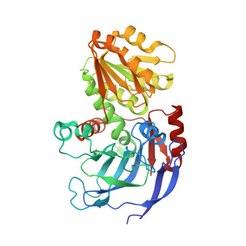
The ternary complex of Pseudomonas aeruginosa alcohol dehydrogenase with NADH and ethylene glycol.

Levin, I.Meiri, G.Peretz, M.Burstein, Y.Frolow, F.

(2004) Protein Sci 13: 1547-1556

  • DOI: https://doi.org/10.1110/ps.03531404
  • Primary Citation Related Structures: 
    1LLU

  • PubMed Abstract: 

    Pseudomonas aeruginosa alcohol dehydrogenase (PaADH; ADH, EC 1.1.1.1) catalyzes the reversible oxidation of primary and secondary alcohols to the corresponding aldehydes and ketones, using NAD as coenzyme. We crystallized the ternary complex of PaADH with its coenzyme and a substrate molecule and determined its structure at a resolution of 2.3 A, using the molecular replacement method. The PaADH tetramer comprises four identical chains of 342 amino acid residues each and obeys ~222-point symmetry. The PaADH monomer is structurally similar to alcohol dehydrogenase monomers from vertebrates, archaea, and bacteria. The stabilization of the ternary complex of PaADH, the coenzyme, and the poor substrate ethylene glycol (k(cat) = 4.5 sec(-1); Km > 200 mM) was due to the blocked exit of the coenzyme in the crystalline state, combined with a high (2.5 M) concentration of the substrate. The structure of the ternary complex presents the precise geometry of the Zn coordination complex, the proton-shuttling system, and the hydride transfer path. The ternary complex structure also suggests that the low efficiency of ethylene glycol as a substrate results from the presence of a second hydroxyl group in this molecule.


  • Organizational Affiliation
    • Department of Organic Chemistry, Weizmann Institute of Science, 76100 Rehovot, Israel.

Macromolecule Content 

  • Total Structure Weight: 294.45 kDa 
  • Atom Count: 21,580 
  • Modeled Residue Count: 2,728 
  • Deposited Residue Count: 2,736 
  • Unique protein chains: 1

Macromolecules

Find similar proteins by:|  3D Structure
Entity ID: 1
MoleculeChains  Sequence LengthOrganismDetailsImage
Alcohol Dehydrogenase
A, B, C, D, E
A, B, C, D, E, F, G, H
342Pseudomonas aeruginosaMutation(s): 0 
Gene Names: adh
EC: 1.1.1.1
UniProt
Find proteins for Q9HTD9 (Pseudomonas aeruginosa (strain ATCC 15692 / DSM 22644 / CIP 104116 / JCM 14847 / LMG 12228 / 1C / PRS 101 / PAO1))
Explore Q9HTD9 
Go to UniProtKB:  Q9HTD9
Entity Groups
Sequence Clusters30% Identity50% Identity70% Identity90% Identity95% Identity100% Identity
UniProt GroupQ9HTD9
Sequence Annotations
Expand
Reference Sequence

Small Molecules

Ligands 3 Unique
IDChains Name / Formula / InChI Key2D Diagram3D Interactions
NAD

Query on NAD


Download:Ideal Coordinates CCD File
CA [auth E]
HA [auth F]
K [auth A]
LA [auth G]
P [auth B]
CA [auth E],
HA [auth F],
K [auth A],
LA [auth G],
P [auth B],
QA [auth H],
T [auth C],
Y [auth D]
NICOTINAMIDE-ADENINE-DINUCLEOTIDE
C21 H27 N7 O14 P2
BAWFJGJZGIEFAR-NNYOXOHSSA-N
ZN

Query on ZN


Download:Ideal Coordinates CCD File
AA [auth E]
BA [auth E]
FA [auth F]
GA [auth F]
I [auth A]
AA [auth E],
BA [auth E],
FA [auth F],
GA [auth F],
I [auth A],
J [auth A],
JA [auth G],
KA [auth G],
N [auth B],
O [auth B],
OA [auth H],
PA [auth H],
R [auth C],
S [auth C],
W [auth D],
X [auth D]
ZINC ION
Zn
PTFCDOFLOPIGGS-UHFFFAOYSA-N
EDO

Query on EDO


Download:Ideal Coordinates CCD File
DA [auth E]
EA [auth E]
IA [auth F]
L [auth A]
M [auth A]
DA [auth E],
EA [auth E],
IA [auth F],
L [auth A],
M [auth A],
MA [auth G],
NA [auth G],
Q [auth B],
RA [auth H],
U [auth C],
V [auth C],
Z [auth D]
1,2-ETHANEDIOL
C2 H6 O2
LYCAIKOWRPUZTN-UHFFFAOYSA-N

Experimental Data & Validation

Experimental Data

  • Method: X-RAY DIFFRACTION
  • Resolution: 2.30 Å
  • R-Value Free:  0.246 (Depositor), 0.249 (DCC) 
  • R-Value Work:  0.229 (Depositor), 0.234 (DCC) 
Space Group: P 1
Unit Cell:
Length ( Å )Angle ( ˚ )
a = 74.171α = 79.34
b = 86.328β = 78.66
c = 125.698γ = 71.58
Software Package:
Software NamePurpose
DENZOdata reduction
SCALEPACKdata scaling
AMoREphasing
MOLREPphasing
CNSrefinement

Structure Validation

View Full Validation Report


Ligand Structure Quality Assessment 


Entry History 

Deposition Data

Revision History  (Full details and data files)

  • Version 1.0: 2003-11-11
    Type: Initial release
  • Version 1.1: 2008-04-28
    Changes: Version format compliance
  • Version 1.2: 2011-07-13
    Changes: Version format compliance
  • Version 1.3: 2023-08-16
    Changes: Data collection, Database references, Derived calculations, Refinement description