1LKI | pdb_00001lki

THE CRYSTAL STRUCTURE AND BIOLOGICAL FUNCTION OF LEUKEMIA INHIBITORY FACTOR: IMPLICATIONS FOR RECEPTOR BINDING


Experimental Data Snapshot

  • Method: X-RAY DIFFRACTION
  • Resolution: 2.00 Å
  • R-Value Work: 
    0.186 (Depositor) 
  • R-Value Observed: 
    0.186 (Depositor) 

wwPDB Validation 3D Report Full Report

Validation slider image for 1LKI

This is version 1.4 of the entry. See complete history

Literature

The crystal structure and biological function of leukemia inhibitory factor: implications for receptor binding.

Robinson, R.C.Grey, L.M.Staunton, D.Vankelecom, H.Vernallis, A.B.Moreau, J.F.Stuart, D.I.Heath, J.K.Jones, E.Y.

(1994) Cell 77: 1101-1116

  • DOI: https://doi.org/10.1016/0092-8674(94)90449-9
  • Primary Citation Related Structures: 
    1LKI

  • PubMed Abstract: 

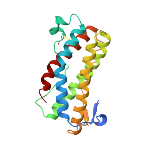
    The structure of murine leukemia inhibitory factor (LIF) has been determined by X-ray crystallography at 2.0 A resolution. The main chain fold conforms to the four alpha-helix bundle topology previously observed for several members of the hematopoietic cytokine family. Of these, LIF shows closest structural homology to granulocyte colony-stimulating factor and growth hormone (GH). Sequence alignments for the functionally related molecules oncostatin M and ciliary neurotrophic factor, when mapped to the LIF structure, indicate regions of conserved surface character. Analysis of the biological function and receptor specificity of a series of human-mouse LIF chimeras implicate two regions of receptor interaction that are located in the fourth helix and the preceding loop. A model for receptor binding based on the structure of the GH ligand-receptor complex requires additional, novel features to account for these data.


  • Organizational Affiliation
    • Department of Biochemistry, University of Oxford, England.

Macromolecule Content 

  • Total Structure Weight: 19.89 kDa 
  • Atom Count: 1,386 
  • Modeled Residue Count: 172 
  • Deposited Residue Count: 180 
  • Unique protein chains: 1

Macromolecules

Find similar proteins by:|  3D Structure
Entity ID: 1
MoleculeChains  Sequence LengthOrganismDetailsImage
LEUKEMIA INHIBITORY FACTOR180Mus musculusMutation(s): 0 
UniProt
Find proteins for P09056 (Mus musculus)
Explore P09056 
Go to UniProtKB:  P09056
Entity Groups
Sequence Clusters30% Identity50% Identity70% Identity90% Identity95% Identity100% Identity
UniProt GroupP09056
Sequence Annotations
Expand
Reference Sequence

Experimental Data & Validation

Experimental Data

  • Method: X-RAY DIFFRACTION
  • Resolution: 2.00 Å
  • R-Value Work:  0.186 (Depositor) 
  • R-Value Observed: 0.186 (Depositor) 
Space Group: P 21 21 21
Unit Cell:
Length ( Å )Angle ( ˚ )
a = 31.1α = 90
b = 56.2β = 90
c = 95.3γ = 90
Software Package:
Software NamePurpose
X-PLORmodel building
X-PLORrefinement
X-PLORphasing

Structure Validation

View Full Validation Report


Entry History 

Deposition Data

Revision History  (Full details and data files)

  • Version 1.0: 1995-03-31
    Type: Initial release
  • Version 1.1: 2008-03-24
    Changes: Version format compliance
  • Version 1.2: 2011-07-13
    Changes: Version format compliance
  • Version 1.3: 2024-06-05
    Changes: Data collection, Database references, Other
  • Version 1.4: 2024-11-20
    Changes: Structure summary