1LK9 | pdb_00001lk9

The Three-dimensional Structure of Alliinase from Garlic


Experimental Data Snapshot

  • Method: X-RAY DIFFRACTION
  • Resolution: 1.53 Å
  • R-Value Free: 
    0.221 (Depositor) 
  • R-Value Work: 
    0.193 (Depositor) 

wwPDB Validation 3D Report Full Report

Validation slider image for 1LK9

Ligand Structure Quality Assessment 


This is version 2.1 of the entry. See complete history

Literature

The active principle of garlic at atomic resolution

Kuettner, E.B.Hilgenfeld, R.Weiss, M.S.

(2002) J Biological Chem 277: 46402-46407

  • DOI: https://doi.org/10.1074/jbc.M208669200
  • Primary Citation Related Structures: 
    1LK9

  • PubMed Abstract: 

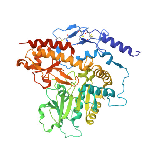
    Despite the fact that many cultures around the world value and utilize garlic as a fundamental component of their cuisine as well as of their medicine cabinets, relatively little is known about the plant's protein configuration that is responsible for the specific properties of garlic. Here, we report the three-dimensional structure of the garlic enzyme alliinase at 1.5 A resolution. Alliinase constitutes the major protein component in garlic bulbs, and it is able to cleave carbon-sulfur bonds. The active enzyme is a pyridoxal-5'-phosphate-dependent homodimeric glycoprotein and belongs to the class I family of pyridoxal-5'-phosphate-dependent enzymes. In addition, it contains a novel epidermal growth factor-like domain that makes it unique among all pyridoxal-5'-phosphate-dependent enzymes.


  • Organizational Affiliation
    • Department of Structural Biology and Crystallography, Institute of Molecular Biotechnology, Beutenbergstrasse 11, D-07745 Jena, Germany.

Macromolecule Content 

  • Total Structure Weight: 107.17 kDa 
  • Atom Count: 7,996 
  • Modeled Residue Count: 852 
  • Deposited Residue Count: 896 
  • Unique protein chains: 1

Macromolecules

Find similar proteins by:|  3D Structure
Entity ID: 1
MoleculeChains  Sequence LengthOrganismDetailsImage
ALLIIN LYASE
A, B
448Allium sativumMutation(s): 0 
EC: 4.4.1.4
UniProt
Find proteins for Q01594 (Allium sativum)
Explore Q01594 
Go to UniProtKB:  Q01594
Entity Groups
Sequence Clusters30% Identity50% Identity70% Identity90% Identity95% Identity100% Identity
UniProt GroupQ01594
Glycosylation
Glycosylation Sites: 2
Sequence Annotations
Expand
Reference Sequence

Oligosaccharides

Help  
Entity ID: 2
MoleculeChains Length2D Diagram GlycosylationD Interactions
beta-D-mannopyranose-(1-4)-2-acetamido-2-deoxy-beta-D-glucopyranose-(1-4)-[alpha-L-fucopyranose-(1-3)]2-acetamido-2-deoxy-beta-D-glucopyranose
C
4N-Glycosylation
Glycosylation Resources
GlyTouCan: G18638YB
GlyCosmos: G18638YB
GlyGen: G18638YB
Entity ID: 3
MoleculeChains Length2D Diagram GlycosylationD Interactions
2-acetamido-2-deoxy-beta-D-glucopyranose-(1-4)-2-acetamido-2-deoxy-beta-D-glucopyranose
D, F
2N-Glycosylation
Glycosylation Resources
GlyTouCan: G42666HT
GlyCosmos: G42666HT
GlyGen: G42666HT
Entity ID: 4
MoleculeChains Length2D Diagram GlycosylationD Interactions
alpha-L-fucopyranose-(1-3)-2-acetamido-2-deoxy-beta-D-glucopyranose
E
2N-Glycosylation
Glycosylation Resources
GlyTouCan: G80587NA
GlyCosmos: G80587NA
GlyGen: G80587NA

Small Molecules

Ligands 5 Unique
IDChains Name / Formula / InChI Key2D Diagram3D Interactions
PLP

Query on PLP


Download:Ideal Coordinates CCD File
N [auth A],
W [auth B]
PYRIDOXAL-5'-PHOSPHATE
C8 H10 N O6 P
NGVDGCNFYWLIFO-UHFFFAOYSA-N
EPE

Query on EPE


Download:Ideal Coordinates CCD File
O [auth A],
P [auth A]
4-(2-HYDROXYETHYL)-1-PIPERAZINE ETHANESULFONIC ACID
C8 H18 N2 O4 S
JKMHFZQWWAIEOD-UHFFFAOYSA-N
SO4

Query on SO4


Download:Ideal Coordinates CCD File
G [auth A]
H [auth A]
I [auth A]
J [auth A]
K [auth A]
G [auth A],
H [auth A],
I [auth A],
J [auth A],
K [auth A],
L [auth A],
Q [auth B],
R [auth B],
S [auth B],
T [auth B],
U [auth B]
SULFATE ION
O4 S
QAOWNCQODCNURD-UHFFFAOYSA-L
DHA

Query on DHA


Download:Ideal Coordinates CCD File
X [auth B]2-AMINO-ACRYLIC ACID
C3 H5 N O2
UQBOJOOOTLPNST-UHFFFAOYSA-N
CL

Query on CL


Download:Ideal Coordinates CCD File
M [auth A],
V [auth B]
CHLORIDE ION
Cl
VEXZGXHMUGYJMC-UHFFFAOYSA-M

Experimental Data & Validation

Experimental Data

  • Method: X-RAY DIFFRACTION
  • Resolution: 1.53 Å
  • R-Value Free:  0.221 (Depositor) 
  • R-Value Work:  0.193 (Depositor) 
Space Group: P 21 21 21
Unit Cell:
Length ( Å )Angle ( ˚ )
a = 68.45α = 90
b = 101.07β = 90
c = 155.69γ = 90
Software Package:
Software NamePurpose
DENZOdata reduction
TRUNCATEdata reduction
MLPHAREphasing
REFMACrefinement
CCP4data scaling

Structure Validation

View Full Validation Report


Ligand Structure Quality Assessment 


Entry History 

Deposition Data

Revision History  (Full details and data files)

  • Version 1.0: 2002-12-11
    Type: Initial release
  • Version 1.1: 2008-04-28
    Changes: Version format compliance
  • Version 1.2: 2011-07-13
    Changes: Non-polymer description, Version format compliance
  • Version 2.0: 2020-07-29
    Type: Remediation
    Reason: Carbohydrate remediation
    Changes: Advisory, Atomic model, Data collection, Derived calculations, Structure summary
  • Version 2.1: 2025-03-26
    Changes: Data collection, Database references, Structure summary