1LI7 | pdb_00001li7

Crystal Structure of Cysteinyl-tRNA Synthetase with Cysteine Substrate Bound


Experimental Data Snapshot

  • Method: X-RAY DIFFRACTION
  • Resolution: 2.60 Å
  • R-Value Free: 
    0.290 (Depositor), 0.289 (DCC) 
  • R-Value Work: 
    0.250 (Depositor), 0.249 (DCC) 
  • R-Value Observed: 
    0.251 (Depositor) 

Starting Model: experimental
View more details

wwPDB Validation 3D Report Full Report

Validation slider image for 1LI7

This is version 1.3 of the entry. See complete history

Literature

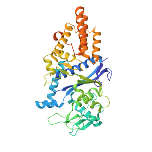
Structural origins of amino acid selection without editing by cysteinyl-tRNA synthetase

Newberry, K.J.Hou, Y.-M.Perona, J.J.

(2002) EMBO J 21: 2778-2787

  • DOI: https://doi.org/10.1093/emboj/21.11.2778
  • Primary Citation Related Structures: 
    1LI5, 1LI7

  • PubMed Abstract: 

    Cysteinyl-tRNA synthetase (CysRS) is highly specific for synthesis of cysteinyl adenylate, yet does not possess the amino acid editing activity characteristic of many other tRNA synthetases. To elucidate how CysRS is able to distinguish cysteine from non-cognate amino acids, crystal structures of the Escherichia coli enzyme were determined in apo and cysteine-bound states. The structures reveal that the substrate cysteine thiolate forms a single direct interaction with a zinc ion bound at the base of the active site cleft, in a trigonal bipyramidal geometry together with four highly conserved protein side chains. Cysteine binding induces movement of the zinc ion towards substrate, as well as flipping of the conserved Trp205 indole ring to pack on the thiol side chain. The imidazole groups of five conserved histidines lie adjacent to the zinc ion, forming a unique arrangement suggestive of functional significance. Thus, amino acid discrimination without editing arises most directly from the favorable zinc-thiolate interaction, which is not possible for non-cognate substrates. Additional selectivity may be generated during the induced-fit conformational changes that help assemble the active site.


  • Organizational Affiliation
    • Department of Chemistry and Biochemistry, Interdepartmental Program in Biomolecular Science and Engineering, University of California at Santa Barbara, Santa Barbara, CA 93106-9510, USA.

Macromolecule Content 

  • Total Structure Weight: 104.92 kDa 
  • Atom Count: 5,827 
  • Modeled Residue Count: 761 
  • Deposited Residue Count: 922 
  • Unique protein chains: 1

Macromolecules

Find similar proteins by:|  3D Structure
Entity ID: 1
MoleculeChains  Sequence LengthOrganismDetailsImage
CYSTEINYL-TRNA SYNTHETASE
A, B
461Escherichia coliMutation(s): 0 
EC: 6.1.1.16
UniProt
Find proteins for P21888 (Escherichia coli (strain K12))
Explore P21888 
Go to UniProtKB:  P21888
Entity Groups
Sequence Clusters30% Identity50% Identity70% Identity90% Identity95% Identity100% Identity
UniProt GroupP21888
Sequence Annotations
Expand
Reference Sequence

Experimental Data & Validation

Experimental Data

  • Method: X-RAY DIFFRACTION
  • Resolution: 2.60 Å
  • R-Value Free:  0.290 (Depositor), 0.289 (DCC) 
  • R-Value Work:  0.250 (Depositor), 0.249 (DCC) 
  • R-Value Observed: 0.251 (Depositor) 
Space Group: P 41 21 2
Unit Cell:
Length ( Å )Angle ( ˚ )
a = 118.98α = 90
b = 118.98β = 90
c = 143.84γ = 90
Software Package:
Software NamePurpose
MOSFLMdata reduction
SCALAdata scaling
CNSrefinement
CCP4data scaling
CNSphasing

Structure Validation

View Full Validation Report


Entry History 

Deposition Data

Revision History  (Full details and data files)

  • Version 1.0: 2002-04-26
    Type: Initial release
  • Version 1.1: 2008-04-28
    Changes: Version format compliance
  • Version 1.2: 2011-07-13
    Changes: Version format compliance
  • Version 1.3: 2023-08-16
    Changes: Data collection, Database references, Derived calculations, Refinement description