1LF1 | pdb_00001lf1

Crystal Structure of Cel5 from Alkalophilic Bacillus sp.


Experimental Data Snapshot

  • Method: X-RAY DIFFRACTION
  • Resolution: 1.70 Å
  • R-Value Free: 
    0.223 (Depositor) 
  • R-Value Work: 
    0.186 (Depositor) 
  • R-Value Observed: 
    0.190 (Depositor) 

wwPDB Validation 3D Report Full Report

Validation slider image for 1LF1

This is version 1.4 of the entry. See complete history

Literature

A novel combination of two classic catalytic schemes.

Shaw, A.Bott, R.Vonrhein, C.Bricogne, G.Power, S.Day, A.G.

(2002) J Mol Biology 320: 303-309

  • DOI: https://doi.org/10.1016/S0022-2836(02)00387-X
  • Primary Citation Related Structures: 
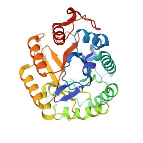
    1LF1

  • PubMed Abstract: 

    The crystal structure of an alkaline Bacillus cellulase catalytic core, from glucoside hydrolase family 5, reveals a novel combination of the catalytic machinery of two classic textbook enzymes. The enzyme has the expected two glutamate residues in close proximity to one another in the active-site that are typical of retaining cellulases. However, the proton donor, glutamate 139 is also unexpectedly a member of a serine-histidine-glutamate catalytic triad, forming a novel combination of catalytic machineries. Structure and sequence analysis of glucoside hydrolase family 5 reveal that the triad is highly conserved, but with variations at the equivalent of the serine position. We speculate that the purpose of this novel catalytic triad is to control the protonation of the acid/base glutamate, facilitating the first step of the catalytic reaction, protonation of the substrate, by the proton donor glutamate. If correct, this will be a novel use for a catalytic triad.


  • Organizational Affiliation
    • Genencor International Inc., 925 Page Mill Road, Palo Alto, CA 94304, USA. ashaw@genencor.com

Macromolecule Content 

  • Total Structure Weight: 34.5 kDa 
  • Atom Count: 2,429 
  • Modeled Residue Count: 296 
  • Deposited Residue Count: 308 
  • Unique protein chains: 1

Macromolecules

Find similar proteins by:|  3D Structure
Entity ID: 1
MoleculeChains  Sequence LengthOrganismDetailsImage
Cel5308Bacillus subtilisMutation(s): 0 
EC: 3.2.1.4
UniProt
Find proteins for A0ACD6B9F0 (Bacillus sp)
Explore A0ACD6B9F0 
Go to UniProtKB:  A0ACD6B9F0
Entity Groups
Sequence Clusters30% Identity50% Identity70% Identity90% Identity95% Identity100% Identity
UniProt GroupA0ACD6B9F0
Sequence Annotations
Expand
Reference Sequence

Experimental Data & Validation

Experimental Data

  • Method: X-RAY DIFFRACTION
  • Resolution: 1.70 Å
  • R-Value Free:  0.223 (Depositor) 
  • R-Value Work:  0.186 (Depositor) 
  • R-Value Observed: 0.190 (Depositor) 
Space Group: P 21 21 21
Unit Cell:
Length ( Å )Angle ( ˚ )
a = 70.44α = 90
b = 78.16β = 90
c = 55.05γ = 90
Software Package:
Software NamePurpose
SHELXL-97refinement

Structure Validation

View Full Validation Report


Entry History 

Deposition Data

Revision History  (Full details and data files)

  • Version 1.0: 2002-07-03
    Type: Initial release
  • Version 1.1: 2008-04-28
    Changes: Version format compliance
  • Version 1.2: 2011-07-13
    Changes: Version format compliance
  • Version 1.3: 2017-10-11
    Changes: Refinement description
  • Version 1.4: 2024-02-14
    Changes: Data collection, Database references