1LEK | pdb_00001lek

Crystal Structure of H-2Kbm3 bound to dEV8


Experimental Data Snapshot

  • Method: X-RAY DIFFRACTION
  • Resolution: 2.15 Å
  • R-Value Free: 
    0.258 (Depositor) 
  • R-Value Work: 
    0.206 (Depositor) 

wwPDB Validation 3D Report Full Report

Validation slider image for 1LEK

This is version 2.1 of the entry. See complete history

Literature

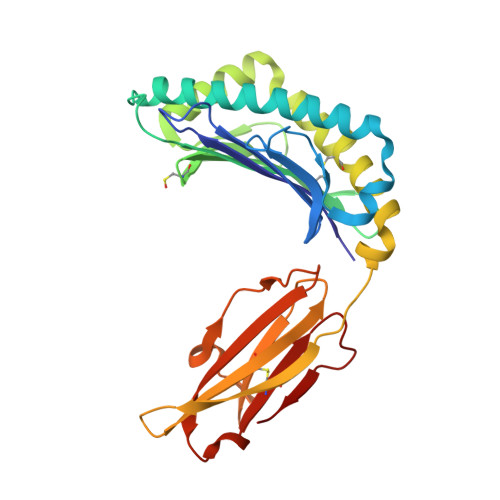
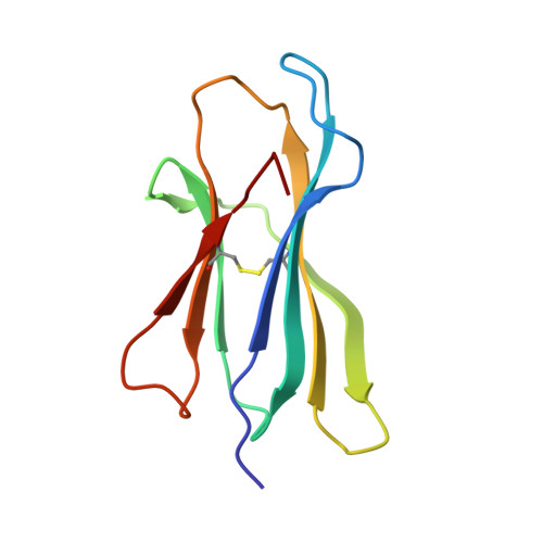
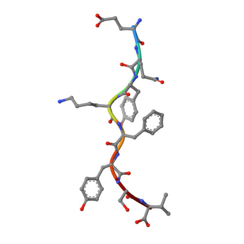
Structural comparison of allogeneic and syngeneic T cell receptor-peptide-major histocompatibility complex complexes: a buried alloreactive mutation subtly alters peptide presentation substantially increasing V(beta) Interactions.

Luz, J.G.Huang, M.Garcia, K.C.Rudolph, M.G.Apostolopoulos, V.Teyton, L.Wilson, I.A.

(2002) J Exp Medicine 195: 1175-1186

  • DOI: https://doi.org/10.1084/jem.20011644
  • Primary Citation Related Structures: 
    1LEG, 1LEK, 1MWA

  • PubMed Abstract: 

    The crystal structures of the 2C/H-2K(bm3)-dEV8 allogeneic complex at 2.4 A and H-2K(bm3)-dEV8 at 2.15 A, when compared with their syngeneic counterparts, elucidate structural changes that induce an alloresponse. The Asp77Ser mutation that imbues H-2K(bm3)-dEV8 with its alloreactive properties is located beneath the peptide and does not directly contact the T cell receptor (TCR). However, the buried mutation induces local rearrangement of the peptide itself to preserve hydrogen bonding interactions between the peptide and the alpha(1) 77 residue. The COOH terminus of the peptide main chain is tugged toward the alpha(1)-helix such that its presentation to the TCR is altered. These changes increase the stability of the allogeneic peptide-major histocompatibility complex (pMHC) complex and increase complementarity in the TCR-pMHC interface, placing greater emphasis on recognition of the pMHC by the TCR beta-chain, evinced by an increase in shape complementarity, buried surface area, and number of TCR-pMHC contacting residues. A nearly fourfold increase in the number of beta-chain-pMHC contacts is accompanied by a concomitant 64% increase in beta-chain-pMHC shape complementarity. Thus, the allogeneic mutation causes the same peptide to be presented differently, temporally and spatially, by the allogeneic and syngeneic MHCs.


  • Organizational Affiliation
    • Department of Molecular Biology, The Scripps Research Institute, La Jolla, CA 92037, USA.

Macromolecule Content 

  • Total Structure Weight: 45.23 kDa 
  • Atom Count: 3,387 
  • Modeled Residue Count: 381 
  • Deposited Residue Count: 381 
  • Unique protein chains: 3

Macromolecules

Find similar proteins by:|  3D Structure
Entity ID: 1
MoleculeChains  Sequence LengthOrganismDetailsImage
H-2 CLASS I HISTOCOMPATIBILITY ANTIGEN, K-B ALPHA CHAIN274Mus musculusMutation(s): 0 
UniProt
Find proteins for P01901 (Mus musculus)
Explore P01901 
Go to UniProtKB:  P01901
Entity Groups
Sequence Clusters30% Identity50% Identity70% Identity90% Identity95% Identity100% Identity
UniProt GroupP01901
Glycosylation
Glycosylation Sites: 2Go to GlyGen: P01901-1
Sequence Annotations
Expand
Reference Sequence
Find similar proteins by:|  3D Structure
Entity ID: 2
MoleculeChains  Sequence LengthOrganismDetailsImage
Beta-2-microglobulin99Mus musculusMutation(s): 0 
UniProt & NIH Common Fund Data Resources
Find proteins for P01887 (Mus musculus)
Explore P01887 
Go to UniProtKB:  P01887
IMPC:  MGI:88127
Entity Groups
Sequence Clusters30% Identity50% Identity70% Identity90% Identity95% Identity100% Identity
UniProt GroupP01887
Sequence Annotations
Expand
Reference Sequence
Find similar proteins by:  Sequence   |   3D Structure  
Entity ID: 3
MoleculeChains  Sequence LengthOrganismDetailsImage
dEV8C [auth P]8N/AMutation(s): 0 
UniProt
Find proteins for Q62425 (Mus musculus)
Explore Q62425 
Go to UniProtKB:  Q62425
Entity Groups
UniProt GroupQ62425
Sequence Annotations
Expand
Reference Sequence

Oligosaccharides

Help  
Entity ID: 4
MoleculeChains Length2D Diagram GlycosylationD Interactions
2-acetamido-2-deoxy-beta-D-glucopyranose-(1-4)-[alpha-L-fucopyranose-(1-6)]2-acetamido-2-deoxy-beta-D-glucopyranoseD [auth C]3N-Glycosylation
Glycosylation Resources
GlyTouCan: G21290RB
GlyCosmos: G21290RB
GlyGen: G21290RB

Small Molecules

Ligands 2 Unique
IDChains Name / Formula / InChI Key2D Diagram3D Interactions
NAG

Query on NAG


Download:Ideal Coordinates CCD File
E [auth A]2-acetamido-2-deoxy-beta-D-glucopyranose
C8 H15 N O6
OVRNDRQMDRJTHS-FMDGEEDCSA-N
PO4

Query on PO4


Download:Ideal Coordinates CCD File
F [auth B]PHOSPHATE ION
O4 P
NBIIXXVUZAFLBC-UHFFFAOYSA-K
Modified Residues  1 Unique
IDChains TypeFormula2D DiagramParent
CSO
Query on CSO
A
L-PEPTIDE LINKINGC3 H7 N O3 SCYS

Experimental Data & Validation

Experimental Data

  • Method: X-RAY DIFFRACTION
  • Resolution: 2.15 Å
  • R-Value Free:  0.258 (Depositor) 
  • R-Value Work:  0.206 (Depositor) 
Space Group: P 21 21 2
Unit Cell:
Length ( Å )Angle ( ˚ )
a = 135.894α = 90
b = 87.123β = 90
c = 45.231γ = 90
Software Package:
Software NamePurpose
DENZOdata reduction
SCALEPACKdata scaling
AMoREphasing
X-PLORrefinement

Structure Validation

View Full Validation Report


Entry History 

Deposition Data

Revision History  (Full details and data files)

  • Version 1.0: 2002-06-26
    Type: Initial release
  • Version 1.1: 2008-04-28
    Changes: Version format compliance
  • Version 1.2: 2011-07-13
    Changes: Non-polymer description, Version format compliance
  • Version 2.0: 2020-07-29
    Type: Remediation
    Reason: Carbohydrate remediation
    Changes: Advisory, Atomic model, Data collection, Derived calculations, Structure summary
  • Version 2.1: 2024-11-13
    Changes: Advisory, Data collection, Database references, Structure summary