1LDN | pdb_00001ldn

STRUCTURE OF A TERNARY COMPLEX OF AN ALLOSTERIC LACTATE DEHYDROGENASE FROM BACILLUS STEAROTHERMOPHILUS AT 2.5 ANGSTROMS RESOLUTION


Experimental Data Snapshot

  • Method: X-RAY DIFFRACTION
  • Resolution: 2.50 Å
  • R-Value Observed: 
    0.157 (Depositor) 

wwPDB Validation 3D Report Full Report

Validation slider image for 1LDN

Ligand Structure Quality Assessment 


This is version 1.4 of the entry. See complete history

Literature

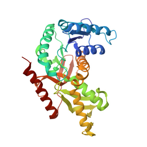
Structure of a ternary complex of an allosteric lactate dehydrogenase from Bacillus stearothermophilus at 2.5 A resolution.

Wigley, D.B.Gamblin, S.J.Turkenburg, J.P.Dodson, E.J.Piontek, K.Muirhead, H.Holbrook, J.J.

(1992) J Mol Biology 223: 317-335

  • DOI: https://doi.org/10.1016/0022-2836(92)90733-z
  • Primary Citation Related Structures: 
    1LDN

  • PubMed Abstract: 

    We report the refined structure of a ternary complex of an allosterically activated lactate dehydrogenase, including the important active site loop. Eightfold non-crystallographic symmetry averaging was utilized to improve the density maps. Interactions between the protein and bound coenzyme and oxamate are described in relation to other studies using site-specific mutagenesis. Fructose 1,6-bisphosphate (FruP2) is bound to the enzyme across one of the 2-fold axes of the tetramer, with the two phosphate moieties interacting with two anion binding sites, one on each of two subunits, across this interface. However, because FruP2 binds at this special site, yet does not possess an internal 2-fold symmetry axis, the ligand is statistically disordered and binds to each site in two different orientations. Binding of FruP2 to the tetramer is signalled to the active site principally through two interactions with His188 and Arg173. His188 is connected to His195 (which binds the carbonyl group of the substrate) and Arg173 is connected to Arg171 (the residue that binds the carboxylate group of the substrate).


  • Organizational Affiliation
    • Department of Biochemistry, University of Leicester, U.K.

Macromolecule Content 

  • Total Structure Weight: 285.34 kDa 
  • Atom Count: 20,716 
  • Modeled Residue Count: 2,528 
  • Deposited Residue Count: 2,528 
  • Unique protein chains: 1

Macromolecules

Find similar proteins by:|  3D Structure
Entity ID: 1
MoleculeChains  Sequence LengthOrganismDetailsImage
L-LACTATE DEHYDROGENASE
A, B, C, D, E
A, B, C, D, E, F, G, H
316Geobacillus stearothermophilusMutation(s): 0 
EC: 1.1.1.27
UniProt
Find proteins for P00344 (Geobacillus stearothermophilus)
Explore P00344 
Go to UniProtKB:  P00344
Entity Groups
Sequence Clusters30% Identity50% Identity70% Identity90% Identity95% Identity100% Identity
UniProt GroupP00344
Sequence Annotations
Expand
Reference Sequence

Small Molecules

Ligands 3 Unique
IDChains Name / Formula / InChI Key2D Diagram3D Interactions
NAD

Query on NAD


Download:Ideal Coordinates CCD File
BA [auth H]
K [auth A]
N [auth B]
P [auth C]
R [auth D]
BA [auth H],
K [auth A],
N [auth B],
P [auth C],
R [auth D],
U [auth E],
X [auth F],
Z [auth G]
NICOTINAMIDE-ADENINE-DINUCLEOTIDE
C21 H27 N7 O14 P2
BAWFJGJZGIEFAR-NNYOXOHSSA-N
FBP

Query on FBP


Download:Ideal Coordinates CCD File
I [auth A],
L [auth B],
S [auth E],
V [auth F]
1,6-di-O-phosphono-beta-D-fructofuranose
C6 H14 O12 P2
RNBGYGVWRKECFJ-ARQDHWQXSA-N
OXM

Query on OXM


Download:Ideal Coordinates CCD File
AA [auth H]
J [auth A]
M [auth B]
O [auth C]
Q [auth D]
AA [auth H],
J [auth A],
M [auth B],
O [auth C],
Q [auth D],
T [auth E],
W [auth F],
Y [auth G]
OXAMIC ACID
C2 H3 N O3
SOWBFZRMHSNYGE-UHFFFAOYSA-N

Experimental Data & Validation

Experimental Data

  • Method: X-RAY DIFFRACTION
  • Resolution: 2.50 Å
  • R-Value Observed: 0.157 (Depositor) 
Space Group: P 1 21 1
Unit Cell:
Length ( Å )Angle ( ˚ )
a = 84.9α = 90
b = 118.2β = 96.07
c = 135.5γ = 90
Software Package:
Software NamePurpose
X-PLORmodel building
PROLSQrefinement
X-PLORrefinement
X-PLORphasing

Structure Validation

View Full Validation Report


Ligand Structure Quality Assessment 


Entry History 

Deposition Data

Revision History  (Full details and data files)

  • Version 1.0: 1994-01-31
    Type: Initial release
  • Version 1.1: 2008-03-24
    Changes: Version format compliance
  • Version 1.2: 2011-07-13
    Changes: Version format compliance
  • Version 1.3: 2020-07-29
    Type: Remediation
    Reason: Carbohydrate remediation
    Changes: Data collection, Derived calculations, Other, Structure summary
  • Version 1.4: 2024-02-14
    Changes: Data collection, Database references, Structure summary