1LCU | pdb_00001lcu

Polylysine Induces an Antiparallel Actin Dimer that Nucleates Filament Assembly: Crystal Structure at 3.5 A Resolution


Experimental Data Snapshot

  • Method: X-RAY DIFFRACTION
  • Resolution: 3.50 Å
  • R-Value Free: 
    0.266 (Depositor) 
  • R-Value Work: 
    0.196 (Depositor) 

Starting Model: experimental
View more details

wwPDB Validation 3D Report Full Report

Validation slider image for 1LCU

This is version 1.5 of the entry. See complete history

Literature

Polylysine induces an antiparallel actin dimer that nucleates filament assembly: crystal structure at 3.5-A resolution.

Bubb, M.R.Govindasamy, L.Yarmola, E.G.Vorobiev, S.M.Almo, S.C.Somasundaram, T.Chapman, M.S.Agbandje-Mckenna, M.Mckenna, R.

(2002) J Biological Chem 277: 20999-21006

  • DOI: https://doi.org/10.1074/jbc.M201371200
  • Primary Citation Related Structures: 
    1IJJ, 1LCU

  • PubMed Abstract: 

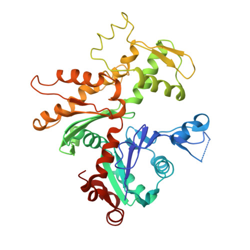
    An antiparallel actin dimer has been proposed to be an intermediate species during actin filament nucleation. We now show that latrunculin A, a marine natural product that inhibits actin polymerization, arrests polylysine-induced nucleation at the level of an antiparallel dimer, resulting in its accumulation. These dimers, when composed of pyrene-labeled actin subunits, give rise to a fluorescent excimer, permitting detection during polymerization in vitro. We report the crystallographic structure of the polylysine-actin-latrunculin A complex at 3.5-A resolution. The non-crystallographic contact is consistent with a dimeric structure and confirms the antiparallel orientation of its subunits. The crystallographic contacts reveal that the mobile DNase I binding loop of one subunit of a symmetry-related antiparallel actin dimer is partially stabilized in the interface between the two subunits of a second antiparallel dimer. These results provide a potential explanation for the paradoxical nucleation of actin filaments that have exclusively parallel subunits by a dimer containing antiparallel subunits.


  • Organizational Affiliation
    • Research Service, Malcom Randall Department of Veterans Affairs Medical Center and the University of Florida College of Medicine, Gainesville, Florida 32608, USA. bubbmr@medicine.ufl.edu

Macromolecule Content 

  • Total Structure Weight: 84.81 kDa 
  • Atom Count: 5,936 
  • Modeled Residue Count: 732 
  • Deposited Residue Count: 742 
  • Unique protein chains: 1

Macromolecules

Find similar proteins by:|  3D Structure
Entity ID: 1
MoleculeChains  Sequence LengthOrganismDetailsImage
Actin, alpha skeletal muscle
A, B
371Oryctolagus cuniculusMutation(s): 0 
EC: 3.6.4
UniProt
Find proteins for P68135 (Oryctolagus cuniculus)
Explore P68135 
Go to UniProtKB:  P68135
Entity Groups
Sequence Clusters30% Identity50% Identity70% Identity90% Identity95% Identity100% Identity
UniProt GroupP68135
Sequence Annotations
Expand
Reference Sequence

Small Molecules

Ligands 4 Unique
IDChains Name / Formula / InChI Key2D Diagram3D Interactions
ATP

Query on ATP


Download:Ideal Coordinates CCD File
H [auth A],
L [auth B]
ADENOSINE-5'-TRIPHOSPHATE
C10 H16 N5 O13 P3
ZKHQWZAMYRWXGA-KQYNXXCUSA-N
LAR

Query on LAR


Download:Ideal Coordinates CCD File
I [auth A],
M [auth B]
LATRUNCULIN A
C22 H31 N O5 S
DDVBPZROPPMBLW-IZGXTMSKSA-N
CA

Query on CA


Download:Ideal Coordinates CCD File
C [auth A],
D [auth A],
F [auth A],
J [auth B]
CALCIUM ION
Ca
BHPQYMZQTOCNFJ-UHFFFAOYSA-N
CL

Query on CL


Download:Ideal Coordinates CCD File
E [auth A],
G [auth A],
K [auth B]
CHLORIDE ION
Cl
VEXZGXHMUGYJMC-UHFFFAOYSA-M

Experimental Data & Validation

Experimental Data

  • Method: X-RAY DIFFRACTION
  • Resolution: 3.50 Å
  • R-Value Free:  0.266 (Depositor) 
  • R-Value Work:  0.196 (Depositor) 
Space Group: P 21 21 21
Unit Cell:
Length ( Å )Angle ( ˚ )
a = 101.456α = 90
b = 103.029β = 90
c = 126.958γ = 90
Software Package:
Software NamePurpose
DENZOdata reduction
SCALEPACKdata scaling
MOLREPphasing
CNSrefinement

Structure Validation

View Full Validation Report


Entry History 

Deposition Data

Revision History  (Full details and data files)

  • Version 1.0: 2002-05-01
    Type: Initial release
  • Version 1.1: 2008-04-28
    Changes: Version format compliance
  • Version 1.2: 2011-07-13
    Changes: Version format compliance
  • Version 1.3: 2017-10-11
    Changes: Refinement description
  • Version 1.4: 2023-08-16
    Changes: Data collection, Database references, Derived calculations, Refinement description
  • Version 1.5: 2024-10-16
    Changes: Structure summary