1L9V | pdb_00001l9v

Non Structural protein encoded by gene segment 8 of rotavirus (NSP2), an NTPase, ssRNA binding and nucleic acid helix-destabilizing protein


Experimental Data Snapshot

  • Method: X-RAY DIFFRACTION
  • Resolution: 2.60 Å
  • R-Value Free: 
    0.287 (Depositor), 0.288 (DCC) 
  • R-Value Work: 
    0.241 (Depositor), 0.242 (DCC) 
  • R-Value Observed: 
    0.241 (Depositor) 

wwPDB Validation 3D Report Full Report

Validation slider image for 1L9V

This is version 1.3 of the entry. See complete history

Literature

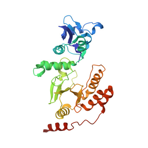
Rotavirus protein involved in genome replication and packaging exhibits a HIT-like fold.

Jayaram, H.Taraporewala, Z.Patton, J.T.Prasad, B.V.

(2002) Nature 417: 311-315

  • DOI: https://doi.org/10.1038/417311a
  • Primary Citation Related Structures: 
    1L9V

  • PubMed Abstract: 

    Rotavirus, the major cause of life-threatening infantile gastroenteritis, is a member of the Reoviridae. Although the structures of rotavirus and other members of the Reoviridae have been extensively studied, little is known about the structures of virus-encoded non-structural proteins that are essential for genome replication and packaging. The non-structural protein NSP2 of rotavirus, which exhibits nucleoside triphosphatase, single-stranded RNA binding, and nucleic-acid helix-destabilizing activities, is a major component of viral replicase complexes. We present here the X-ray structure of the functional octamer of NSP2 determined to a resolution of 2.6 A. The NSP2 monomer has two distinct domains. The amino-terminal domain has a new fold. The carboxy-terminal domain resembles the ubiquitous cellular histidine triad (HIT) group of nucleotidyl hydrolases. This structural similarity suggests that the nucleotide-binding site is located inside the cleft between the two domains. Prominent grooves that run diagonally across the doughnut-shaped octamer are probable locations for RNA binding. Several RNA binding sites, resulting from the quaternary organization of NSP2 monomers, may be required for the helix destabilizing activity of NSP2 and its function during genome replication and packaging.


  • Organizational Affiliation
    • Program in Structural and Computational Biology and Molecular Biophysics, Verna and Marrs McLean Department of Biochemistry, Baylor College of Medicine, Houston, Texas 77030, USA.

Macromolecule Content 

  • Total Structure Weight: 36.62 kDa 
  • Atom Count: 2,546 
  • Modeled Residue Count: 313 
  • Deposited Residue Count: 317 
  • Unique protein chains: 1

Macromolecules

Find similar proteins by:|  3D Structure
Entity ID: 1
MoleculeChains  Sequence LengthOrganismDetailsImage
Rotavirus-NSP2317Simian 11 rotavirus (serotype 3 / strain SA11-Ramig)Mutation(s): 0 
Gene Names: gene segment 8
EC: 3.6.4
UniProt
Find proteins for Q03243 (Rotavirus A (strain RVA/SA11-Ramig/G3P[X]))
Explore Q03243 
Go to UniProtKB:  Q03243
Entity Groups
Sequence Clusters30% Identity50% Identity70% Identity90% Identity95% Identity100% Identity
UniProt GroupQ03243
Sequence Annotations
Expand
Reference Sequence

Experimental Data & Validation

Experimental Data

  • Method: X-RAY DIFFRACTION
  • Resolution: 2.60 Å
  • R-Value Free:  0.287 (Depositor), 0.288 (DCC) 
  • R-Value Work:  0.241 (Depositor), 0.242 (DCC) 
  • R-Value Observed: 0.241 (Depositor) 
Space Group: I 4 2 2
Unit Cell:
Length ( Å )Angle ( ˚ )
a = 108.432α = 90
b = 108.432β = 90
c = 152.877γ = 90
Software Package:
Software NamePurpose
CNSrefinement
DENZOdata reduction
SCALEPACKdata scaling
CNSphasing

Structure Validation

View Full Validation Report


Entry History 

Deposition Data

Revision History  (Full details and data files)

  • Version 1.0: 2002-06-05
    Type: Initial release
  • Version 1.1: 2008-04-28
    Changes: Version format compliance
  • Version 1.2: 2011-07-13
    Changes: Derived calculations, Version format compliance
  • Version 1.3: 2024-02-14
    Changes: Data collection, Database references