1L2S | pdb_00001l2s

X-ray crystal structure of AmpC beta-lactamase from E. coli in complex with a DOCK-predicted non-covalent inhibitor


Experimental Data Snapshot

  • Method: X-RAY DIFFRACTION
  • Resolution: 1.94 Å
  • R-Value Free: 
    0.207 (Depositor), 0.204 (DCC) 
  • R-Value Work: 
    0.173 (Depositor), 0.173 (DCC) 

Starting Model: experimental
View more details

wwPDB Validation 3D Report Full Report

Validation slider image for 1L2S

Ligand Structure Quality Assessment 


This is version 1.3 of the entry. See complete history

Literature

Structure-based discovery of a novel, noncovalent inhibitor of AmpC beta-lactamase.

Powers, R.A.Morandi, F.Shoichet, B.K.

(2002) Structure 10: 1013-1023

  • DOI: https://doi.org/10.1016/s0969-2126(02)00799-2
  • Primary Citation Related Structures: 
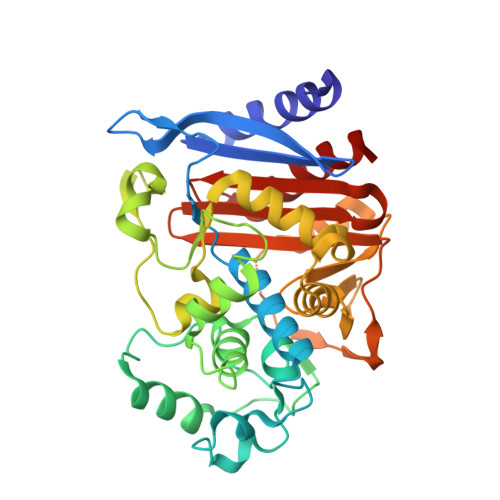
    1L2S

  • PubMed Abstract: 

    beta-lactamases are the most widespread resistance mechanisms to beta-lactam antibiotics, and there is a pressing need for novel, non-beta-lactam drugs. A database of over 200,000 compounds was docked to the active site of AmpC beta-lactamase to identify potential inhibitors. Fifty-six compounds were tested, and three had K(i) values of 650 microM or better. The best of these, 3-[(4-chloroanilino)sulfonyl]thiophene-2-carboxylic acid, was a competitive noncovalent inhibitor (K(i) = 26 microM), which also reversed resistance to beta-lactams in bacteria expressing AmpC. The structure of AmpC in complex with this compound was determined by X-ray crystallography to 1.94 A and reveals that the inhibitor interacts with key active-site residues in sites targeted in the docking calculation. Indeed, the experimentally determined conformation of the inhibitor closely resembles the prediction. The structure of the enzyme-inhibitor complex presents an opportunity to improve binding affinity in a novel series of inhibitors discovered by structure-based methods.


  • Organizational Affiliation
    • Department of Molecular Pharmacology and Biological Chemistry, Northwestern University, 303 East Chicago Avenue, IL 60611, USA.

Macromolecule Content 

  • Total Structure Weight: 80.13 kDa 
  • Atom Count: 5,916 
  • Modeled Residue Count: 713 
  • Deposited Residue Count: 716 
  • Unique protein chains: 1

Macromolecules

Find similar proteins by:|  3D Structure
Entity ID: 1
MoleculeChains  Sequence LengthOrganismDetailsImage
beta-lactamase
A, B
358Escherichia coliMutation(s): 0 
Gene Names: K12
EC: 3.5.2.6
UniProt
Find proteins for P00811 (Escherichia coli (strain K12))
Explore P00811 
Go to UniProtKB:  P00811
Entity Groups
Sequence Clusters30% Identity50% Identity70% Identity90% Identity95% Identity100% Identity
UniProt GroupP00811
Sequence Annotations
Expand
Reference Sequence

Small Molecules

Binding Affinity Annotations 
IDSourceBinding Affinity
STC BindingDB:  1L2S Ki: 2.60e+4 (nM) from 1 assay(s)

Experimental Data & Validation

Experimental Data

  • Method: X-RAY DIFFRACTION
  • Resolution: 1.94 Å
  • R-Value Free:  0.207 (Depositor), 0.204 (DCC) 
  • R-Value Work:  0.173 (Depositor), 0.173 (DCC) 
Space Group: C 1 2 1
Unit Cell:
Length ( Å )Angle ( ˚ )
a = 118.67α = 90
b = 76.42β = 116.63
c = 97.9γ = 90
Software Package:
Software NamePurpose
CNSrefinement
DENZOdata reduction
SCALEPACKdata scaling
CNSphasing

Structure Validation

View Full Validation Report


Ligand Structure Quality Assessment 


Entry History 

Deposition Data

Revision History  (Full details and data files)

  • Version 1.0: 2002-07-17
    Type: Initial release
  • Version 1.1: 2008-04-28
    Changes: Version format compliance
  • Version 1.2: 2011-07-13
    Changes: Version format compliance
  • Version 1.3: 2023-08-16
    Changes: Data collection, Database references, Derived calculations, Refinement description