The structure of a replication initiator unites diverse aspects of nucleic acid metabolism
Campos-Olivas, R., Louis, J.M., Clerot, D., Gronenborn, B., Gronenborn, A.M.(2002) Proc Natl Acad Sci U S A 99: 10310-10315
- PubMed: 12130667 Search on PubMedSearch on PubMed Central
- DOI: https://doi.org/10.1073/pnas.152342699
- Primary Citation Related Structures:
1L2M, 1L5I - PubMed Abstract:
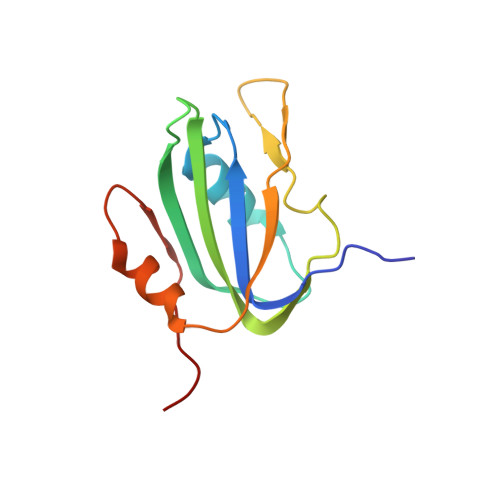
Rolling circle replication is a mechanism for copying single-stranded genomes by means of double-stranded intermediates. A multifunctional replication initiator protein (Rep) is indispensable for the precise initiation and termination of this process. Despite the ubiquitous presence and fundamental importance of rolling circle replication elements, structural information on their respective replication initiators is still missing. Here we present the solution NMR structure of the catalytic domain of Rep, the initiator protein of tomato yellow leaf curl virus. It is composed of a central five-stranded anti-parallel beta-sheet, flanked by a small two-stranded beta-sheet, a beta-hairpin and two alpha-helices. Surprisingly, the structure reveals that the catalytic Rep domain is related to a large group of proteins that bind RNA or DNA. Identification of Rep as resembling the family of ribonucleoprotein/RNA-recognition motif fold proteins establishes a structure-based evolutionary link between RNA binding proteins, splicing factors, and replication initiators of prokaryotic and eukaryotic single-stranded DNA elements and mammalian DNA tumor viruses.
- Laboratory of Chemical Physics, National Institute of Diabetes and Digestive and Kidney Diseases, National Institutes of Health, Bethesda, MD 20892, USA.
Organizational Affiliation: