1L2A | pdb_00001l2a

The Crystal Structure and Catalytic Mechanism of Cellobiohydrolase CelS, the Major Enzymatic Component of the Clostridium thermocellum cellulosome


Experimental Data Snapshot

  • Method: X-RAY DIFFRACTION
  • Resolution: 2.50 Å
  • R-Value Free: 
    0.226 (Depositor), 0.223 (DCC) 
  • R-Value Work: 
    0.181 (Depositor), 0.181 (DCC) 
  • R-Value Observed: 
    0.183 (Depositor) 

wwPDB Validation 3D Report Full Report

Validation slider image for 1L2A

This is version 2.1 of the entry. See complete history

Literature

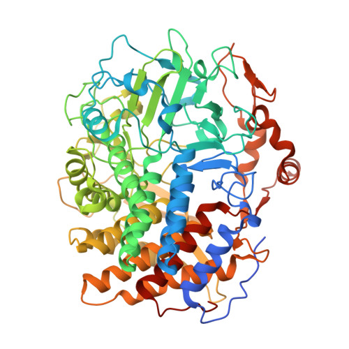
The crystal structure and catalytic mechanism of cellobiohydrolase CelS, the major enzymatic component of the Clostridium thermocellum Cellulosome.

Guimaraes, B.G.Souchon, H.Lytle, B.L.David Wu, J.H.Alzari, P.M.

(2002) J Mol Biology 320: 587-596

  • DOI: https://doi.org/10.1016/s0022-2836(02)00497-7
  • Primary Citation Related Structures: 
    1L1Y, 1L2A

  • PubMed Abstract: 

    Cellobiohydrolase CelS plays an important role in the cellulosome, an active cellulase system produced by the thermophilic anaerobe Clostridium thermocellum. The structures of the catalytic domain of CelS in complex with substrate (cellohexaose) and product (cellobiose) were determined at 2.5 and 2.4 A resolution, respectively. The protein folds into an (alpha/alpha)(6) barrel with a tunnel-shaped substrate-binding region. The conformation of the loops defining the tunnel is intrinsically stable in the absence of substrate, suggesting a model to account for the processive mode of action of family 48 cellobiohydrolases. Structural comparisons with other (alpha/alpha)(6) barrel glycosidases indicate that CelS and endoglucanase CelA, a sequence-unrelated family 8 glycosidase with a groove-shaped substrate-binding region, use the same catalytic machinery to hydrolyze the glycosidic linkage, despite a low sequence similarity and a different endo/exo mode of action. A remarkable feature of the mechanism is the absence, from CelS, of a carboxylic group acting as the base catalyst. The nearly identical arrangement of substrate and functionally important residues in the two active sites strongly suggests an evolutionary relationship between the cellobiohydrolase and endoglucanase families, which can therefore be classified into a new clan of glycoside hydrolases.


  • Organizational Affiliation
    • Unité de Biochimie Structurale, CNRS URA 2185, Institut Pasteur, 25 rue du Dr. Roux, 75724 Paris cedex 15, France.

Macromolecule Content 

  • Total Structure Weight: 467.83 kDa 
  • Atom Count: 32,414 
  • Modeled Residue Count: 3,852 
  • Deposited Residue Count: 4,068 
  • Unique protein chains: 1

Macromolecules

Find similar proteins by:|  3D Structure
Entity ID: 1
MoleculeChains  Sequence LengthOrganismDetailsImage
cellobiohydrolase
A, B, C, D, E
A, B, C, D, E, F
678Acetivibrio thermocellusMutation(s): 0 
Gene Names: cels
EC: 3.2.1.4 (PDB Primary Data), 3.2.1.176 (UniProt)
UniProt
Find proteins for P0C2S5 (Acetivibrio thermocellus)
Explore P0C2S5 
Go to UniProtKB:  P0C2S5
Entity Groups
Sequence Clusters30% Identity50% Identity70% Identity90% Identity95% Identity100% Identity
UniProt GroupP0C2S5
Sequence Annotations
Expand
Reference Sequence

Oligosaccharides

Help  
Entity ID: 2
MoleculeChains Length2D Diagram GlycosylationD Interactions
beta-D-glucopyranose-(1-4)-beta-D-glucopyranose
G, I, K, M, O
G, I, K, M, O, Q
2N/A
Glycosylation Resources
GlyTouCan: G84824ZO
GlyCosmos: G84824ZO
Entity ID: 3
MoleculeChains Length2D Diagram GlycosylationD Interactions
beta-D-glucopyranose-(1-4)-beta-D-glucopyranose-(1-4)-beta-D-glucopyranose-(1-4)-beta-D-glucopyranose-(1-4)-beta-D-glucopyranose-(1-4)-beta-D-glucopyranose
H, J, L, N, P
H, J, L, N, P, R
6N/A
Glycosylation Resources
GlyTouCan: G09454VW
GlyCosmos: G09454VW
GlyGen: G09454VW

Experimental Data & Validation

Experimental Data

  • Method: X-RAY DIFFRACTION
  • Resolution: 2.50 Å
  • R-Value Free:  0.226 (Depositor), 0.223 (DCC) 
  • R-Value Work:  0.181 (Depositor), 0.181 (DCC) 
  • R-Value Observed: 0.183 (Depositor) 
Space Group: P 21 21 21
Unit Cell:
Length ( Å )Angle ( ˚ )
a = 148.027α = 90
b = 207.64β = 90
c = 215.354γ = 90
Software Package:
Software NamePurpose
REFMACrefinement
DENZOdata reduction
SCALEPACKdata scaling

Structure Validation

View Full Validation Report


Entry History 

Deposition Data

Revision History  (Full details and data files)

  • Version 1.0: 2002-07-17
    Type: Initial release
  • Version 1.1: 2008-04-28
    Changes: Version format compliance
  • Version 1.2: 2011-07-13
    Changes: Version format compliance
  • Version 2.0: 2020-07-29
    Type: Remediation
    Reason: Carbohydrate remediation
    Changes: Advisory, Atomic model, Data collection, Derived calculations, Structure summary
  • Version 2.1: 2024-02-14
    Changes: Data collection, Database references, Structure summary