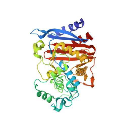
Structural bases of stability-function tradeoffs in enzymes.
Beadle, B.M., Shoichet, B.K.(2002) J Mol Biol 321: 285-296
- PubMed: 12144785
- DOI: https://doi.org/10.1016/s0022-2836(02)00599-5
- Primary Citation of Related Structures:
1L0D, 1L0E, 1L0F, 1L0G - PubMed Abstract:
The structures of enzymes reflect two tendencies that appear opposed. On one hand, they fold into compact, stable structures; on the other hand, they bind a ligand and catalyze a reaction. To be stable, enzymes fold to maximize favorable interactions, forming a tightly packed hydrophobic core, exposing hydrophilic groups, and optimizing intramolecular hydrogen-bonding. To be functional, enzymes carve out an active site for ligand binding, exposing hydrophobic surface area, clustering like charges, and providing unfulfilled hydrogen bond donors and acceptors. Using AmpC beta-lactamase, an enzyme that is well-characterized structurally and mechanistically, the relationship between enzyme stability and function was investigated by substituting key active-site residues and measuring the changes in stability and activity. Substitutions of catalytic residues Ser64, Lys67, Tyr150, Asn152, and Lys315 decrease the activity of the enzyme by 10(3)-10(5)-fold compared to wild-type. Concomitantly, many of these substitutions increase the stability of the enzyme significantly, by up to 4.7kcal/mol. To determine the structural origins of stabilization, the crystal structures of four mutant enzymes were determined to between 1.90A and 1.50A resolution. These structures revealed several mechanisms by which stability was increased, including mimicry of the substrate by the substituted residue (S64D), relief of steric strain (S64G), relief of electrostatic strain (K67Q), and improved polar complementarity (N152H). These results suggest that the preorganization of functionality characteristic of active sites has come at a considerable cost to enzyme stability. In proteins of unknown function, the presence of such destabilized regions may indicate the presence of a binding site.
Organizational Affiliation:
Department of Molecular Pharmacology and Biological Chemistry, Northwestern University School of Medicine, 303 East Chicago Avenue S215, Chicago, IL 60611-3008, USA.


