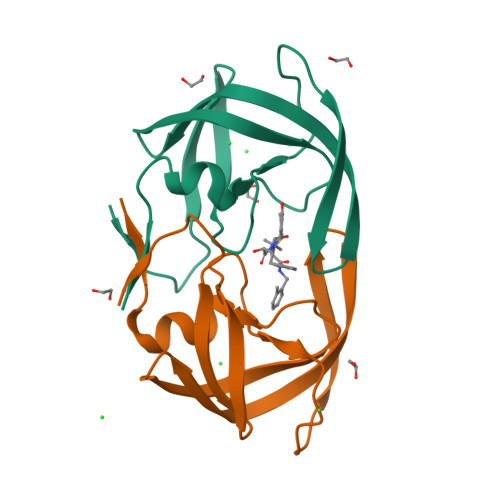
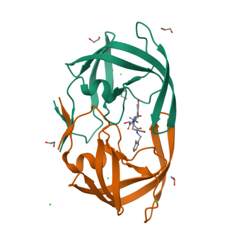
1KZK

JE-2147-HIV Protease Complex


Experimental Data Snapshot

  • Method: X-RAY DIFFRACTION
  • Resolution: 1.09 Å
  • R-Value Free: 0.189 
  • R-Value Work: 0.151 
  • R-Value Observed: 0.152 

Starting Model: experimental
View more details

wwPDB Validation   3D Report Full Report


Ligand Structure Quality Assessment 


This is version 1.6 of the entry. See complete history


Literature

Anisotropic Dynamics of the JE-2147-HIV Protease Complex: Drug Resistance and Thermodynamic Binding Mode Examined in a 1.09 A Structure

Reiling, K.K.Endres, N.F.Dauber, D.S.Craik, C.S.Stroud, R.M.

(2002) Biochemistry 41: 4582-4594

  • DOI: https://doi.org/10.1021/bi011781z
  • Primary Citation of Related Structures:  
    1KZK

  • PubMed Abstract: 

    The structure of HIV protease (HIV Pr) bound to JE-2147 (also named AG1776 or KNI-764) is determined here to 1.09 A resolution. This highest-resolution structure for HIV Pr allows refinement of anisotropic displacement parameters (ADPs) for all atoms. Clustering based on the directional information in ADPs defines two sets of subdomains such that within each set, subdomains undergo similar anisotropic motion. These sets are (a) the core of monomer A grouped with both substrate-binding flaps and (b) the core of monomer B coupled to both catalytic aspartates (25A/B). The four-stranded beta-sheet (1-4 A/B and 95-99 A/B) that forms a significant part of the dimer interface exhibits large anisotropic amplitudes that differ from those of the other sets of subdomains. JE-2147 is shown here to be a picomolar inhibitor (K(i) = 41 +/- 18 pM). The structure is used to interpret the mechanism of association of JE-2147, a second-generation inhibitor for which binding is enthalpically driven, with respect to first-generation inhibitors for which binding is predominantly entropically driven [Velazquez-Campoy, A., et al. (2001) Arch. Biochem. Biophys. 390, 169-175]. Relative to the entropically driven inhibitor complexes, the JE-2147-HIV Pr complex exhibits an approximately 0.5 A movement of the substrate flaps in toward the substrate, suggesting a more compatible enthalpically driven association. Domains of the protease identified by clustering of ADPs also suggest a model of enthalpy-entropy compensation for all HIV Pr inhibitors in which dynamic coupling of the flaps is offset by an increased level of motion of the beta-sheet domain of the dimer interface (1-4 A/B and 95-99 A/B).


  • Organizational Affiliation

    Department of Biochemistry and Biophysics, Graduate Group in Biophysics, University of California at San Francisco, San Francisco, California 94143, USA.


Macromolecules
Find similar proteins by:  (by identity cutoff)  |  3D Structure
Entity ID: 1
MoleculeChains Sequence LengthOrganismDetailsImage
Protease
A, B
99Human immunodeficiency virusMutation(s): 1 
EC: 3.4.23.16
UniProt
Find proteins for P03369 (Human immunodeficiency virus type 1 group M subtype B (isolate ARV2/SF2))
Explore P03369 
Go to UniProtKB:  P03369
Entity Groups  
Sequence Clusters30% Identity50% Identity70% Identity90% Identity95% Identity100% Identity
UniProt GroupP03369
Sequence Annotations
Expand
  • Reference Sequence
Small Molecules
Ligands 3 Unique
IDChains Name / Formula / InChI Key2D Diagram3D Interactions
JE2
Query on JE2

Download Ideal Coordinates CCD File 
C [auth A](4R)-3-{(2S,3S)-2-hydroxy-3-[(3-hydroxy-2-methylbenzoyl)amino]-4-phenylbutanoyl}-5,5-dimethyl-N-(2-methylbenzyl)-1,3-thiazolidine-4-carboxamide
C32 H37 N3 O5 S
CUFQBQOBLVLKRF-RZDMPUFOSA-N
EDO
Query on EDO

Download Ideal Coordinates CCD File 
F [auth A],
G [auth A],
H [auth A],
I [auth A],
M [auth B]
1,2-ETHANEDIOL
C2 H6 O2
LYCAIKOWRPUZTN-UHFFFAOYSA-N
CL
Query on CL

Download Ideal Coordinates CCD File 
D [auth A],
E [auth A],
J [auth B],
K [auth B],
L [auth B]
CHLORIDE ION
Cl
VEXZGXHMUGYJMC-UHFFFAOYSA-M
Binding Affinity Annotations 
IDSourceBinding Affinity
JE2 PDBBind:  1KZK Ki: 0.04 (nM) from 1 assay(s)
Biologically Interesting Molecules (External Reference) 1 Unique
Entity ID: 2
IDChains NameType/Class2D Diagram3D Interactions
PRD_000566 (JE2)
Query on PRD_000566
C [auth A]KNI-764Peptide-like / Enzyme inhibitor
Experimental Data & Validation

Experimental Data

  • Method: X-RAY DIFFRACTION
  • Resolution: 1.09 Å
  • R-Value Free: 0.189 
  • R-Value Work: 0.151 
  • R-Value Observed: 0.152 
  • Space Group: P 21 21 21
Unit Cell:
Length ( Å )Angle ( ˚ )
a = 51.203α = 90
b = 58.409β = 90
c = 62.858γ = 90
Software Package:
Software NamePurpose
AMoREphasing
SHELXL-97refinement
DENZOdata reduction
SCALEPACKdata scaling

Structure Validation

View Full Validation Report


Ligand Structure Quality Assessment 


Entry History 

Deposition Data

Revision History  (Full details and data files)

  • Version 1.0: 2002-04-03
    Type: Initial release
  • Version 1.1: 2008-04-28
    Changes: Version format compliance
  • Version 1.2: 2011-07-13
    Changes: Atomic model, Database references, Derived calculations, Non-polymer description, Structure summary, Version format compliance
  • Version 1.3: 2012-12-12
    Changes: Other
  • Version 1.4: 2018-01-24
    Changes: Database references
  • Version 1.5: 2021-10-27
    Changes: Database references, Derived calculations, Structure summary
  • Version 1.6: 2023-08-16
    Changes: Data collection, Refinement description