1KZF | pdb_00001kzf

Crystal Structure of the Acyl-homoserine Lactone Synthase, EsaI


Experimental Data Snapshot

  • Method: X-RAY DIFFRACTION
  • Resolution: 1.80 Å
  • R-Value Free: 
    0.244 (Depositor) 
  • R-Value Work: 
    0.209 (Depositor) 
  • R-Value Observed: 
    0.224 (Depositor) 

Starting Model: experimental
View more details

wwPDB Validation 3D Report Full Report

Validation slider image for 1KZF

This is version 1.3 of the entry. See complete history

Literature

Structural basis and specificity of acyl-homoserine lactone signal production in bacterial quorum sensing.

Watson, W.T.Minogue, T.D.Val, D.L.von Bodman, S.B.Churchill, M.E.

(2002) Mol Cell 9: 685-694

  • DOI: https://doi.org/10.1016/s1097-2765(02)00480-x
  • Primary Citation Related Structures: 
    1K4J, 1KZF

  • PubMed Abstract: 

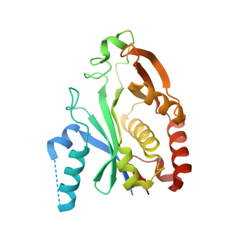
    Synthesis and detection of acyl-homoserine lactones (AHLs) enables many gram-negative bacteria to engage in quorum sensing, an intercellular signaling mechanism that activates differentiation to virulent and biofilm lifestyles. The AHL synthases catalyze acylation of S-adenosyl-L-methionine by acyl-acyl carrier protein and lactonization of the methionine moiety to give AHLs. The crystal structure of the AHL synthase, EsaI, determined at 1.8 A resolution, reveals a remarkable structural similarity to the N-acetyltransferases and defines a common phosphopantetheine binding fold as the catalytic core. Critical residues responsible for catalysis and acyl chain specificity have been identified from a modeled substrate complex and verified through functional analysis in vivo. A mechanism for the N-acylation of S-adenosyl-L-methionine by 3-oxo-hexanoyl-acyl carrier protein is proposed.


  • Organizational Affiliation
    • Department of Pharmacology, The University of Colorado Health Sciences Center, 4200 E. Ninth Avenue, Denver, CO 80262, USA.

Macromolecule Content 

  • Total Structure Weight: 26.04 kDa 
  • Atom Count: 1,649 
  • Modeled Residue Count: 198 
  • Deposited Residue Count: 230 
  • Unique protein chains: 1

Macromolecules

Find similar proteins by:|  3D Structure
Entity ID: 1
MoleculeChains  Sequence LengthOrganismDetailsImage
acyl-homoserinelactone synthase EsaI230Pantoea stewartii subsp. stewartiiMutation(s): 0 
Gene Names: EsaI/EsaR gene cluster
EC: 2.3.1.184
UniProt
Find proteins for P54656 (Pantoea stewartii subsp. stewartii)
Explore P54656 
Go to UniProtKB:  P54656
Entity Groups
Sequence Clusters30% Identity50% Identity70% Identity90% Identity95% Identity100% Identity
UniProt GroupP54656
Sequence Annotations
Expand
Reference Sequence

Experimental Data & Validation

Experimental Data

  • Method: X-RAY DIFFRACTION
  • Resolution: 1.80 Å
  • R-Value Free:  0.244 (Depositor) 
  • R-Value Work:  0.209 (Depositor) 
  • R-Value Observed: 0.224 (Depositor) 
Space Group: P 43
Unit Cell:
Length ( Å )Angle ( ˚ )
a = 66.991α = 90
b = 66.991β = 90
c = 47.011γ = 90
Software Package:
Software NamePurpose
CNSrefinement
DENZOdata reduction
SCALEPACKdata scaling
CNSphasing

Structure Validation

View Full Validation Report


Entry History 

Deposition Data

Revision History  (Full details and data files)

  • Version 1.0: 2002-04-17
    Type: Initial release
  • Version 1.1: 2008-04-28
    Changes: Version format compliance
  • Version 1.2: 2011-07-13
    Changes: Version format compliance
  • Version 1.3: 2023-08-16
    Changes: Data collection, Database references, Refinement description