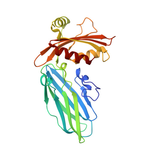
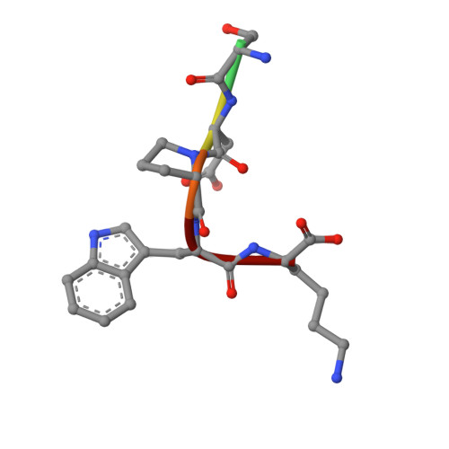
1KYD

AP-2 CLATHRIN ADAPTOR ALPHA-APPENDAGE IN COMPLEX WITH EPSIN DPW PEPTIDE


Experimental Data Snapshot

  • Method: X-RAY DIFFRACTION
  • Resolution: 2.00 Å
  • R-Value Free: 0.228 
  • R-Value Work: 0.178 

Starting Model: experimental
View more details

wwPDB Validation   3D Report Full Report


This is version 1.4 of the entry. See complete history


Literature

Accessory protein recruitment motifs in clathrin-mediated endocytosis.

Brett, T.J.Traub, L.M.Fremont, D.H.

(2002) Structure 10: 797-809

  • DOI: https://doi.org/10.1016/s0969-2126(02)00784-0
  • Primary Citation of Related Structures:  
    1KY6, 1KY7, 1KYD, 1KYF, 1KYU

  • PubMed Abstract: 

    Clathrin-mediated endocytosis depends upon the interaction of accessory proteins with the alpha-ear of the AP-2 adaptor. We present structural characterization of these regulatory interactions. DPF and DPW motif peptides derived from eps15 and epsin bind in type I beta turn conformations to a conserved pocket on the alpha-ear platform. We show evidence for a second binding site that is DPW motif specific. The structure of a complex with an AP-2 binding segment from amphiphysin reveals a novel binding motif that we term FxDxF, which is engaged in an extended conformation by a unique surface of the platform domain. The FxDxF motif is also used by AP180 and the 170 kDa isoform of synaptojanin and can be found in several potential endocytic proteins, including HIP1, CD2AP, and PLAP. A mechanism of clathrin assembly regulation is suggested by three different AP-2 engagement modes.


  • Organizational Affiliation

    Department of Pathology and Immunology, Washington University School of Medicine, St. Louis, MO 63110, USA.


Macromolecules
Find similar proteins by:  (by identity cutoff)  |  3D Structure
Entity ID: 1
MoleculeChains Sequence LengthOrganismDetailsImage
ALPHA-ADAPTIN C247Mus musculusMutation(s): 0 
UniProt & NIH Common Fund Data Resources
Find proteins for P17427 (Mus musculus)
Explore P17427 
Go to UniProtKB:  P17427
IMPC:  MGI:101920
Entity Groups  
Sequence Clusters30% Identity50% Identity70% Identity90% Identity95% Identity100% Identity
UniProt GroupP17427
Sequence Annotations
Expand
  • Reference Sequence

Find similar proteins by:  Sequence   |   3D Structure  

Entity ID: 2
MoleculeChains Sequence LengthOrganismDetailsImage
EH domain-binding mitotic phosphoproteinB [auth P]6N/AMutation(s): 1 
UniProt & NIH Common Fund Data Resources
Find proteins for Q9Y6I3 (Homo sapiens)
Explore Q9Y6I3 
Go to UniProtKB:  Q9Y6I3
PHAROS:  Q9Y6I3
GTEx:  ENSG00000063245 
Entity Groups  
UniProt GroupQ9Y6I3
Sequence Annotations
Expand
  • Reference Sequence
Small Molecules
Ligands 1 Unique
IDChains Name / Formula / InChI Key2D Diagram3D Interactions
SO4
Query on SO4

Download Ideal Coordinates CCD File 
C [auth A]SULFATE ION
O4 S
QAOWNCQODCNURD-UHFFFAOYSA-L
Experimental Data & Validation

Experimental Data

  • Method: X-RAY DIFFRACTION
  • Resolution: 2.00 Å
  • R-Value Free: 0.228 
  • R-Value Work: 0.178 
  • Space Group: P 1 21 1
Unit Cell:
Length ( Å )Angle ( ˚ )
a = 40.78α = 90
b = 71.88β = 99.97
c = 41.67γ = 90
Software Package:
Software NamePurpose
CNSrefinement
DENZOdata reduction
SCALEPACKdata scaling
CNSphasing

Structure Validation

View Full Validation Report


Entry History 

Deposition Data

Revision History  (Full details and data files)

  • Version 1.0: 2002-06-12
    Type: Initial release
  • Version 1.1: 2008-04-28
    Changes: Version format compliance
  • Version 1.2: 2011-07-13
    Changes: Version format compliance
  • Version 1.3: 2021-10-27
    Changes: Database references, Derived calculations
  • Version 1.4: 2023-08-16
    Changes: Data collection, Refinement description