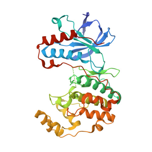
1KV1

p38 MAP Kinase in Complex with Inhibitor 1


Experimental Data Snapshot

  • Method: X-RAY DIFFRACTION
  • Resolution: 2.50 Å
  • R-Value Free: 0.284 
  • R-Value Work: 0.221 
  • R-Value Observed: 0.230 

wwPDB Validation   3D Report Full Report


This is version 1.3 of the entry. See complete history


Literature

Inhibition of p38 MAP kinase by utilizing a novel allosteric binding site.

Pargellis, C.Tong, L.Churchill, L.Cirillo, P.F.Gilmore, T.Graham, A.G.Grob, P.M.Hickey, E.R.Moss, N.Pav, S.Regan, J.

(2002) Nat Struct Biol 9: 268-272

  • DOI: https://doi.org/10.1038/nsb770
  • Primary Citation of Related Structures:  
    1KV1, 1KV2

  • PubMed Abstract: 

    The p38 MAP kinase plays a crucial role in regulating the production of proinflammatory cytokines, such as tumor necrosis factor and interleukin-1. Blocking this kinase may offer an effective therapy for treating many inflammatory diseases. Here we report a new allosteric binding site for a diaryl urea class of highly potent and selective inhibitors against human p38 MAP kinase. The formation of this binding site requires a large conformational change not observed previously for any of the protein Ser/Thr kinases. This change is in the highly conserved Asp-Phe-Gly motif within the active site of the kinase. Solution studies demonstrate that this class of compounds has slow binding kinetics, consistent with the requirement for conformational change. Improving interactions in this allosteric pocket, as well as establishing binding interactions in the ATP pocket, enhanced the affinity of the inhibitors by 12,000-fold. One of the most potent compounds in this series, BIRB 796, has picomolar affinity for the kinase and low nanomolar inhibitory activity in cell culture.


  • Organizational Affiliation

    Department of Biology, Boehringer Ingelheim Pharmaceuticals, Research and Development Center, 900 Ridgebury Road, Ridgefield, Connecticut 06877, USA. cpargel@rdg.boehringer-ingelheim.com


Macromolecules
Find similar proteins by:  (by identity cutoff)  |  3D Structure
Entity ID: 1
MoleculeChains Sequence LengthOrganismDetailsImage
p38 MAP kinase360Homo sapiensMutation(s): 0 
EC: 2.7.1 (PDB Primary Data), 2.7.11.24 (UniProt)
UniProt & NIH Common Fund Data Resources
Find proteins for Q16539 (Homo sapiens)
Explore Q16539 
Go to UniProtKB:  Q16539
PHAROS:  Q16539
GTEx:  ENSG00000112062 
Entity Groups  
Sequence Clusters30% Identity50% Identity70% Identity90% Identity95% Identity100% Identity
UniProt GroupQ16539
Sequence Annotations
Expand
  • Reference Sequence
Small Molecules
Ligands 1 Unique
IDChains Name / Formula / InChI Key2D Diagram3D Interactions
BMU
Query on BMU

Download Ideal Coordinates CCD File 
B [auth A]1-(5-TERT-BUTYL-2-METHYL-2H-PYRAZOL-3-YL)-3-(4-CHLORO-PHENYL)-UREA
C15 H19 Cl N4 O
FWIJKWMXNHRSRO-UHFFFAOYSA-N
Binding Affinity Annotations 
IDSourceBinding Affinity
BMU BindingDB:  1KV1 Kd: min: 1160, max: 1190 (nM) from 2 assay(s)
IC50: min: 794, max: 2.10e+4 (nM) from 3 assay(s)
PDBBind:  1KV1 Kd: 1160 (nM) from 1 assay(s)
Experimental Data & Validation

Experimental Data

  • Method: X-RAY DIFFRACTION
  • Resolution: 2.50 Å
  • R-Value Free: 0.284 
  • R-Value Work: 0.221 
  • R-Value Observed: 0.230 
  • Space Group: P 21 21 21
Unit Cell:
Length ( Å )Angle ( ˚ )
a = 65α = 90
b = 74.4β = 90
c = 77.1γ = 90
Software Package:
Software NamePurpose
CNSrefinement
DENZOdata reduction
SCALEPACKdata scaling

Structure Validation

View Full Validation Report


Entry History 

Deposition Data

Revision History  (Full details and data files)

  • Version 1.0: 2002-03-27
    Type: Initial release
  • Version 1.1: 2008-04-27
    Changes: Version format compliance
  • Version 1.2: 2011-07-13
    Changes: Version format compliance
  • Version 1.3: 2024-02-14
    Changes: Data collection, Database references, Derived calculations