1KU6 | pdb_00001ku6

Fasciculin 2-Mouse Acetylcholinesterase Complex


Experimental Data Snapshot

  • Method: X-RAY DIFFRACTION
  • Resolution: 2.50 Å
  • R-Value Free: 
    0.273 (Depositor), 0.280 (DCC) 
  • R-Value Work: 
    0.226 (Depositor), 0.230 (DCC) 

wwPDB Validation 3D Report Full Report

Validation slider image for 1KU6

Ligand Structure Quality Assessment 


This is version 1.4 of the entry. See complete history

Literature

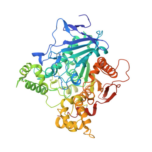
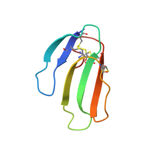
Structural insights into ligand interactions at the acetylcholinesterase peripheral anionic site.

Bourne, Y.Taylor, P.Radic, Z.Marchot, P.

(2003) EMBO J 22: 1-12

  • DOI: https://doi.org/10.1093/emboj/cdg005
  • Primary Citation Related Structures: 
    1J06, 1J07, 1KU6, 1N5M, 1N5R

  • PubMed Abstract: 

    The peripheral anionic site on acetylcholinesterase (AChE), located at the active center gorge entry, encompasses overlapping binding sites for allosteric activators and inhibitors; yet, the molecular mechanisms coupling this site to the active center at the gorge base to modulate catalysis remain unclear. The peripheral site has also been proposed to be involved in heterologous protein associations occurring during synaptogenesis or upon neurodegeneration. A novel crystal form of mouse AChE, combined with spectrophotometric analyses of the crystals, enabled us to solve unique structures of AChE with a free peripheral site, and as three complexes with peripheral site inhibitors: the phenylphenanthridinium ligands, decidium and propidium, and the pyrogallol ligand, gallamine, at 2.20-2.35 A resolution. Comparison with structures of AChE complexes with the peptide fasciculin or with organic bifunctional inhibitors unveils new structural determinants contributing to ligand interactions at the peripheral site, and permits a detailed topographic delineation of this site. Hence, these structures provide templates for designing compounds directed to the enzyme surface that modulate specific surface interactions controlling catalytic activity and non-catalytic heterologous protein associations.


  • Organizational Affiliation
    • Architecture et Fonction des Macromolécules Biologiques, CNRS UMR 6098, Marseille, France. yves@afmb.cnrs-mrs.fr

Macromolecule Content 

  • Total Structure Weight: 67.46 kDa 
  • Atom Count: 4,824 
  • Modeled Residue Count: 596 
  • Deposited Residue Count: 610 
  • Unique protein chains: 2

Macromolecules

Find similar proteins by:|  3D Structure
Entity ID: 1
MoleculeChains  Sequence LengthOrganismDetailsImage
ACETYLCHOLINESTERASE549Mus musculusMutation(s): 0 
EC: 3.1.1.7
UniProt & NIH Common Fund Data Resources
Find proteins for P21836 (Mus musculus)
Explore P21836 
Go to UniProtKB:  P21836
IMPC:  MGI:87876
Entity Groups
Sequence Clusters30% Identity50% Identity70% Identity90% Identity95% Identity100% Identity
UniProt GroupP21836
Glycosylation
Glycosylation Sites: 1Go to GlyGen: P21836-1
Sequence Annotations
Expand
Reference Sequence
Find similar proteins by:|  3D Structure
Entity ID: 2
MoleculeChains  Sequence LengthOrganismDetailsImage
FASCICULIN 261Dendroaspis angusticepsMutation(s): 0 
UniProt
Find proteins for P0C1Z0 (Dendroaspis angusticeps)
Explore P0C1Z0 
Go to UniProtKB:  P0C1Z0
Entity Groups
Sequence Clusters30% Identity50% Identity70% Identity90% Identity95% Identity100% Identity
UniProt GroupP0C1Z0
Sequence Annotations
Expand
Reference Sequence

Small Molecules

Ligands 2 Unique
IDChains Name / Formula / InChI Key2D Diagram3D Interactions
NAG

Query on NAG


Download:Ideal Coordinates CCD File
C [auth A]2-acetamido-2-deoxy-beta-D-glucopyranose
C8 H15 N O6
OVRNDRQMDRJTHS-FMDGEEDCSA-N
EDO

Query on EDO


Download:Ideal Coordinates CCD File
D [auth A]1,2-ETHANEDIOL
C2 H6 O2
LYCAIKOWRPUZTN-UHFFFAOYSA-N

Experimental Data & Validation

Experimental Data

  • Method: X-RAY DIFFRACTION
  • Resolution: 2.50 Å
  • R-Value Free:  0.273 (Depositor), 0.280 (DCC) 
  • R-Value Work:  0.226 (Depositor), 0.230 (DCC) 
Space Group: P 65 2 2
Unit Cell:
Length ( Å )Angle ( ˚ )
a = 73.8α = 90
b = 73.8β = 90
c = 548.62γ = 120
Software Package:
Software NamePurpose
DENZOdata reduction
SCALAdata scaling
CNSrefinement
CCP4data scaling
CNSphasing

Structure Validation

View Full Validation Report


Ligand Structure Quality Assessment 


Entry History 

Deposition Data

Revision History  (Full details and data files)

  • Version 1.0: 2003-12-23
    Type: Initial release
  • Version 1.1: 2008-04-27
    Changes: Version format compliance
  • Version 1.2: 2011-07-13
    Changes: Non-polymer description, Version format compliance
  • Version 1.3: 2020-07-29
    Type: Remediation
    Reason: Carbohydrate remediation
    Changes: Advisory, Data collection, Derived calculations, Structure summary
  • Version 1.4: 2024-10-30
    Changes: Advisory, Data collection, Database references, Structure summary