1KU0 | pdb_00001ku0

Structure of the Bacillus stearothermophilus L1 lipase


Experimental Data Snapshot

  • Method: X-RAY DIFFRACTION
  • Resolution: 2.00 Å
  • R-Value Free: 
    0.218 (Depositor), 0.217 (DCC) 
  • R-Value Work: 
    0.185 (Depositor), 0.185 (DCC) 

wwPDB Validation 3D Report Full Report

Validation slider image for 1KU0

This is version 1.4 of the entry. See complete history

Literature

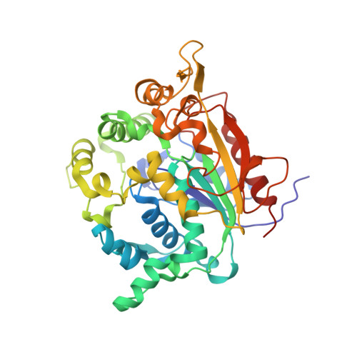
Novel zinc-binding center and a temperature switch in the Bacillus stearothermophilus L1 lipase.

Jeong, S.T.Kim, H.K.Kim, S.J.Chi, S.W.Pan, J.G.Oh, T.K.Ryu, S.E.

(2002) J Biological Chem 277: 17041-17047

  • DOI: https://doi.org/10.1074/jbc.M200640200
  • Primary Citation Related Structures: 
    1KU0

  • PubMed Abstract: 

    The bacterial thermoalkalophilic lipases optimally hydrolyze saturated fatty acids at elevated temperatures. They also have significant sequence homology with staphylococcal lipases, and both the thermoalkalophilic and staphylococcal lipases are grouped as the lipase family I.5. We report here the first crystal structure of the lipase family I.5, the structure of a thermoalkalophilic lipase from Bacillus stearothermophilus L1 (L1 lipase) determined at 2.0-A resolution. The structure is in a closed conformation, and the active site is buried under a long lid helix. Unexpectedly, the structure exhibits a zinc-binding site in an extra domain that accounts for the larger molecular size of the family I.5 enzymes in comparison to other microbial lipases. The zinc-coordinated extra domain makes tight interactions with the loop extended from the C terminus of the lid helix, suggesting that the activation of the family I.5 lipases may be regulated by the strength of the interactions. The unusually long lid helix makes strong hydrophobic interactions with its neighbors. The structural information together with previous biochemical observations indicate that the temperature-mediated lid opening is triggered by the thermal dissociation of the hydrophobic interactions.


  • Organizational Affiliation
    • Center for Cellular Switch Protein Structure and Environmental Bioresources Lab, Korea Research Institute of Bioscience and Biotechnology, 52 Euh-eun-dong, Yusong-gu, Daejon, Korea.

Macromolecule Content 

  • Total Structure Weight: 86.6 kDa 
  • Atom Count: 6,347 
  • Modeled Residue Count: 773 
  • Deposited Residue Count: 776 
  • Unique protein chains: 1

Macromolecules

Find similar proteins by:|  3D Structure
Entity ID: 1
MoleculeChains  Sequence LengthOrganismDetailsImage
L1 lipase
A, B
388Geobacillus stearothermophilusMutation(s): 0 
EC: 3.1.1.3
UniProt
Find proteins for O66015 (Geobacillus stearothermophilus)
Explore O66015 
Go to UniProtKB:  O66015
Entity Groups
Sequence Clusters30% Identity50% Identity70% Identity90% Identity95% Identity100% Identity
UniProt GroupO66015
Sequence Annotations
Expand
Reference Sequence

Experimental Data & Validation

Experimental Data

  • Method: X-RAY DIFFRACTION
  • Resolution: 2.00 Å
  • R-Value Free:  0.218 (Depositor), 0.217 (DCC) 
  • R-Value Work:  0.185 (Depositor), 0.185 (DCC) 
Space Group: C 1 2 1
Unit Cell:
Length ( Å )Angle ( ˚ )
a = 119.62α = 90
b = 85.04β = 99.73
c = 98.36γ = 90
Software Package:
Software NamePurpose
SHARPphasing
CNSrefinement

Structure Validation

View Full Validation Report


Entry History 

Deposition Data

Revision History  (Full details and data files)

  • Version 1.0: 2002-08-21
    Type: Initial release
  • Version 1.1: 2008-04-01
    Changes: Version format compliance
  • Version 1.2: 2011-07-13
    Changes: Version format compliance
  • Version 1.3: 2017-10-11
    Changes: Refinement description
  • Version 1.4: 2024-03-13
    Changes: Data collection, Database references, Derived calculations