1KTB | pdb_00001ktb

The Structure of alpha-N-Acetylgalactosaminidase


Experimental Data Snapshot

  • Method: X-RAY DIFFRACTION
  • Resolution: 1.90 Å
  • R-Value Free: 
    0.222 (Depositor), 0.218 (DCC) 
  • R-Value Work: 
    0.211 (Depositor), 0.212 (DCC) 
  • R-Value Observed: 
    0.211 (Depositor) 

wwPDB Validation 3D Report Full Report

Validation slider image for 1KTB

Ligand Structure Quality Assessment 


This is version 2.1 of the entry. See complete history

Literature

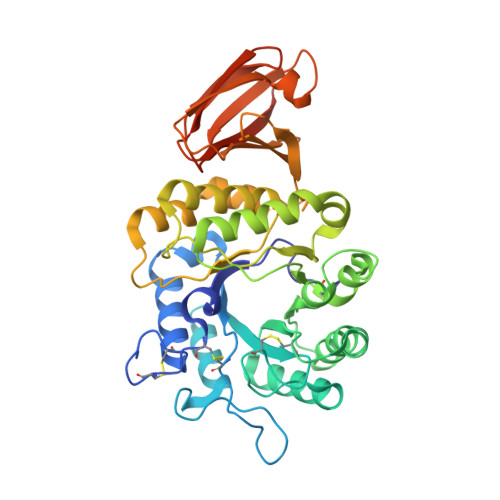
The 1.9 A structure of alpha-N-acetylgalactosaminidase: molecular basis of glycosidase deficiency diseases

Garman, S.C.Hannick, L.Zhu, A.Garboczi, D.N.

(2002) Structure 10: 425-434

  • DOI: https://doi.org/10.1016/s0969-2126(02)00726-8
  • Primary Citation Related Structures: 
    1KTB, 1KTC

  • PubMed Abstract: 

    In the lysosome, glycosidases degrade glycolipids, glycoproteins, and oligosaccharides. Mutations in glycosidases cause disorders characterized by the deposition of undegraded carbohydrates. Schindler and Fabry diseases are caused by the incomplete degradation of carbohydrates with terminal alpha-N-acetylgalactosamine and alpha-galactose, respectively. Here we present the X-ray structure of alpha-N-acetylgalactosaminidase (alpha-NAGAL), the glycosidase that removes alpha-N-acetylgalactosamine, and the structure with bound ligand. The active site residues of alpha-NAGAL are conserved in the closely related enzyme a-galactosidase A (alpha-GAL). The structure demonstrates the catalytic mechanisms of both enzymes and reveals the structural basis of mutations causing Schindler and Fabry diseases. As alpha-NAGAL and alpha-GAL produce type O "universal donor" blood from type A and type B blood, the alpha-NAGAL structure will aid in the engineering of improved enzymes for blood conversion.


  • Organizational Affiliation
    • Structural Biology Section, Laboratory of Immunogenetics, National Institute of Allergy and Infectious Diseases, National Institutes of Health, Rockville, Maryland 20852, USA. garman@alpha.niaid.nih.gov

Macromolecule Content 

  • Total Structure Weight: 48.16 kDa 
  • Atom Count: 3,637 
  • Modeled Residue Count: 388 
  • Deposited Residue Count: 405 
  • Unique protein chains: 1

Macromolecules

Find similar proteins by:|  3D Structure
Entity ID: 1
MoleculeChains  Sequence LengthOrganismDetailsImage
alpha-N-acetylgalactosaminidase405Gallus gallusMutation(s): 0 
Gene Names: alpha-NAGA
EC: 3.2.1.49
UniProt
Find proteins for Q90744 (Gallus gallus)
Explore Q90744 
Go to UniProtKB:  Q90744
Entity Groups
Sequence Clusters30% Identity50% Identity70% Identity90% Identity95% Identity100% Identity
UniProt GroupQ90744
Glycosylation
Glycosylation Sites: 3
Sequence Annotations
Expand
Reference Sequence

Oligosaccharides

Help  
Entity ID: 2
MoleculeChains Length2D Diagram GlycosylationD Interactions
2-acetamido-2-deoxy-beta-D-glucopyranose-(1-4)-2-acetamido-2-deoxy-beta-D-glucopyranose
B
2N-Glycosylation
Glycosylation Resources
GlyTouCan: G42666HT
GlyCosmos: G42666HT
GlyGen: G42666HT

Small Molecules

Ligands 4 Unique
IDChains Name / Formula / InChI Key2D Diagram3D Interactions
NAG

Query on NAG


Download:Ideal Coordinates CCD File
C [auth A],
D [auth A]
2-acetamido-2-deoxy-beta-D-glucopyranose
C8 H15 N O6
OVRNDRQMDRJTHS-FMDGEEDCSA-N
SO4

Query on SO4


Download:Ideal Coordinates CCD File
E [auth A],
F [auth A],
G [auth A],
H [auth A],
I [auth A]
SULFATE ION
O4 S
QAOWNCQODCNURD-UHFFFAOYSA-L
GOL

Query on GOL


Download:Ideal Coordinates CCD File
J [auth A]
K [auth A]
L [auth A]
M [auth A]
N [auth A]
J [auth A],
K [auth A],
L [auth A],
M [auth A],
N [auth A],
O [auth A],
P [auth A],
Q [auth A],
R [auth A],
S [auth A],
T [auth A],
U [auth A]
GLYCEROL
C3 H8 O3
PEDCQBHIVMGVHV-UHFFFAOYSA-N
ACY

Query on ACY


Download:Ideal Coordinates CCD File
V [auth A],
W [auth A]
ACETIC ACID
C2 H4 O2
QTBSBXVTEAMEQO-UHFFFAOYSA-N

Experimental Data & Validation

Experimental Data

  • Method: X-RAY DIFFRACTION
  • Resolution: 1.90 Å
  • R-Value Free:  0.222 (Depositor), 0.218 (DCC) 
  • R-Value Work:  0.211 (Depositor), 0.212 (DCC) 
  • R-Value Observed: 0.211 (Depositor) 
Space Group: P 43 21 2
Unit Cell:
Length ( Å )Angle ( ˚ )
a = 72.593α = 90
b = 72.593β = 90
c = 176.066γ = 90
Software Package:
Software NamePurpose
DENZOdata reduction
SCALEPACKdata scaling
CNSrefinement
CNSphasing

Structure Validation

View Full Validation Report


Ligand Structure Quality Assessment 


Entry History 

Deposition Data

Revision History  (Full details and data files)

  • Version 1.0: 2002-03-15
    Type: Initial release
  • Version 1.1: 2008-04-27
    Changes: Version format compliance
  • Version 1.2: 2011-07-13
    Changes: Non-polymer description, Version format compliance
  • Version 1.3: 2011-11-16
    Changes: Atomic model
  • Version 2.0: 2020-07-29
    Type: Remediation
    Reason: Carbohydrate remediation
    Changes: Atomic model, Data collection, Database references, Derived calculations, Structure summary
  • Version 2.1: 2024-11-20
    Changes: Data collection, Database references, Structure summary