1KRC | pdb_00001krc

CRYSTAL STRUCTURE OF KLEBSIELLA AEROGENES UREASE, ITS APOENZYME AND TWO ACTIVE SITE MUTANTS


Experimental Data Snapshot

  • Method: X-RAY DIFFRACTION
  • Resolution: 2.50 Å
  • R-Value Work: 
    0.180 (Depositor), 0.167 (DCC) 
  • R-Value Observed: 
    0.180 (Depositor) 

wwPDB Validation 3D Report Full Report

Validation slider image for 1KRC

This is version 1.3 of the entry. See complete history

Literature

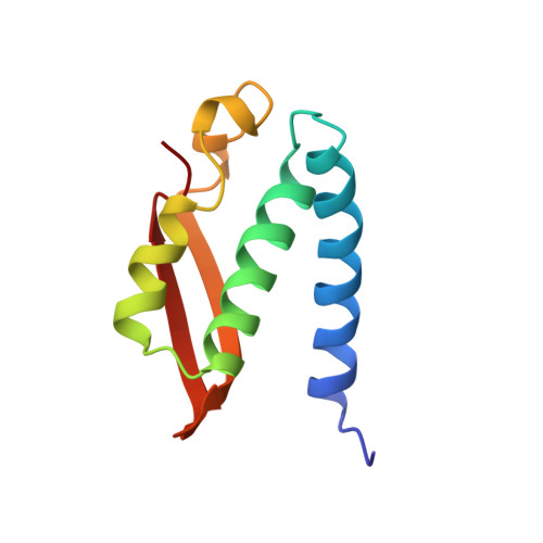
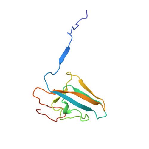
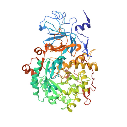
Structures of the Klebsiella aerogenes urease apoenzyme and two active-site mutants.

Jabri, E.Karplus, P.A.

(1996) Biochemistry 35: 10616-10626

  • DOI: https://doi.org/10.1021/bi960424z
  • Primary Citation Related Structures: 
    1KRA, 1KRB, 1KRC

  • PubMed Abstract: 

    Urease from Klebsiella aerogenes [Jabri et al. (1995) Science 268, 998-1004] is an (alpha beta gamma)3 trimer with each alpha-subunit having an (alpha beta)8-barrel domain containing a binickel active center. Here we examine structure-function relations for urease in more detail through structural analysis of the urease apoenzyme at 2.3 A resolution and mutants of two key catalytic residues (H219A and H320A) at 2.5 A resolution. With the exception of the active site, in which a water molecule takes the place of the missing carbamate and nickel atoms, the structure of the apoenzyme is nearly identical to that of the holoenzyme, suggesting a high degree of preorganization which helps explain the tight binding of nickel. In the structure of H219A, the major change involves a conformational shift and ordering of the active site flap, but a small shift in the side chain of Asp alpha 221 could contribute to the lower activity of H219A. In the H320A structure, the catalytic water, primarily a Ni-2 ligand in the holoenzyme, shifts into a bridging position. This shift shows that the nickel ligation is rather sensitive to the environment and the change in ligation may contribute to the 10(5)-fold lower activity of H320A. In addition, these results show that urease is resilient to the loss of nickel ions and mutations. Analysis of the urease tertiary/quaternary structure suggests that the stability of this enzyme may be largely due to its burial of an unusually large fraction of its residues: 50% in the gamma-subunit, 30% in the beta-subunit, and 60% in the alpha-subunit.


  • Organizational Affiliation
    • Section of Biochemistry, Molecular and Cell Biology, Cornell University, Ithaca, New York 14853, USA.

Macromolecule Content 

  • Total Structure Weight: 83.27 kDa 
  • Atom Count: 5,943 
  • Modeled Residue Count: 767 
  • Deposited Residue Count: 773 
  • Unique protein chains: 3

Macromolecules

Find similar proteins by:|  3D Structure
Entity ID: 1
MoleculeChains  Sequence LengthOrganismDetailsImage
UREASE100Klebsiella aerogenesMutation(s): 0 
EC: 3.5.1.5
UniProt
Find proteins for P18316 (Klebsiella aerogenes)
Explore P18316 
Go to UniProtKB:  P18316
Entity Groups
Sequence Clusters30% Identity50% Identity70% Identity90% Identity95% Identity100% Identity
UniProt GroupP18316
Sequence Annotations
Expand
Reference Sequence
Find similar proteins by:|  3D Structure
Entity ID: 2
MoleculeChains  Sequence LengthOrganismDetailsImage
UREASE106Klebsiella aerogenesMutation(s): 0 
EC: 3.5.1.5
UniProt
Find proteins for P18315 (Klebsiella aerogenes)
Explore P18315 
Go to UniProtKB:  P18315
Entity Groups
Sequence Clusters30% Identity50% Identity70% Identity90% Identity95% Identity100% Identity
UniProt GroupP18315
Sequence Annotations
Expand
Reference Sequence
Find similar proteins by:|  3D Structure
Entity ID: 3
MoleculeChains  Sequence LengthOrganismDetailsImage
UREASE567Klebsiella aerogenesMutation(s): 0 
EC: 3.5.1.5
UniProt
Find proteins for P18314 (Klebsiella aerogenes)
Explore P18314 
Go to UniProtKB:  P18314
Entity Groups
Sequence Clusters30% Identity50% Identity70% Identity90% Identity95% Identity100% Identity
UniProt GroupP18314
Sequence Annotations
Expand
Reference Sequence

Experimental Data & Validation

Experimental Data

  • Method: X-RAY DIFFRACTION
  • Resolution: 2.50 Å
  • R-Value Work:  0.180 (Depositor), 0.167 (DCC) 
  • R-Value Observed: 0.180 (Depositor) 
Space Group: I 21 3
Unit Cell:
Length ( Å )Angle ( ˚ )
a = 170.8α = 90
b = 170.8β = 90
c = 170.8γ = 90
Software Package:
Software NamePurpose
X-PLORmodel building
X-PLORrefinement
DENZOdata reduction
SCALEPACKdata scaling
X-PLORphasing

Structure Validation

View Full Validation Report


Entry History 

Deposition Data

Revision History  (Full details and data files)

  • Version 1.0: 1995-10-15
    Type: Initial release
  • Version 1.1: 2008-03-03
    Changes: Version format compliance
  • Version 1.2: 2011-07-13
    Changes: Derived calculations, Version format compliance
  • Version 1.3: 2024-06-05
    Changes: Data collection, Database references, Derived calculations, Other