1KR5

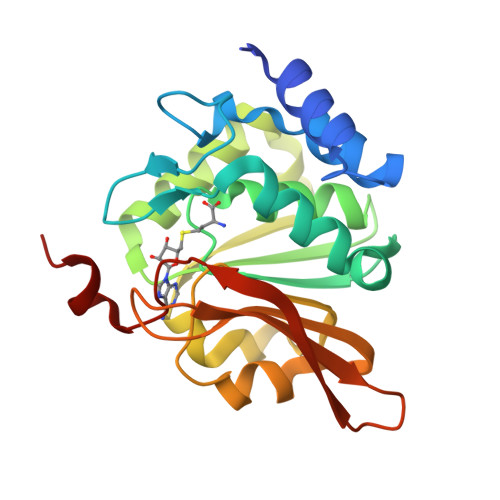
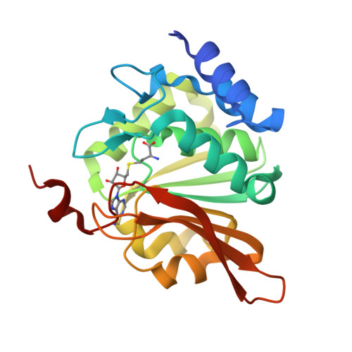
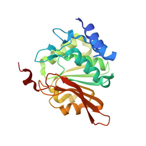
Crystal structure of human L-isoaspartyl methyltransferase


Experimental Data Snapshot

  • Method: X-RAY DIFFRACTION
  • Resolution: 2.10 Å
  • R-Value Free: 0.264 
  • R-Value Work: 0.221 
  • R-Value Observed: 0.224 

Starting Model: experimental
View more details

wwPDB Validation   3D Report Full Report


Ligand Structure Quality Assessment 


This is version 1.4 of the entry. See complete history


Literature

Crystal structure of human L-isoaspartyl methyltransferase.

Ryttersgaard, C.Griffith, S.C.Sawaya, M.R.MacLaren, D.C.Clarke, S.Yeates, T.O.

(2002) J Biol Chem 277: 10642-10646

  • DOI: https://doi.org/10.1074/jbc.M200229200
  • Primary Citation of Related Structures:  
    1KR5

  • PubMed Abstract: 

    The enzyme l-isoaspartyl methyltransferase initiates the repair of damaged proteins by recognizing and methylating isomerized and racemized aspartyl residues in aging proteins. The crystal structure of the human enzyme containing a bound S-adenosyl-l-homocysteine cofactor is reported here at a resolution of 2.1 A. A comparison of the human enzyme to homologs from two other species reveals several significant differences among otherwise similar structures. In all three structures, we find that three conserved charged residues are buried in the protein interior near the active site. Electrostatics calculations suggest that these buried charges might make significant contributions to the energetics of binding the charged S-adenosyl-l-methionine cofactor and to catalysis. We suggest a possible structural explanation for the observed differences in reactivity toward the structurally similar l-isoaspartyl and d-aspartyl residues in the human, archael, and eubacterial enzymes. Finally, the human structure reveals that the known genetic polymorphism at residue 119 (Val/Ile) maps to an exposed region away from the active site.


  • Organizational Affiliation

    Department of Chemistry and Biochemistry, University of California, Los Angeles, California 90095-1569, USA.


Macromolecules
Find similar proteins by:  (by identity cutoff)  |  3D Structure
Entity ID: 1
MoleculeChains Sequence LengthOrganismDetailsImage
Protein-L-isoaspartate O-methyltransferase226Homo sapiensMutation(s): 1 
EC: 2.1.1.77
UniProt & NIH Common Fund Data Resources
Find proteins for P22061 (Homo sapiens)
Explore P22061 
Go to UniProtKB:  P22061
PHAROS:  P22061
GTEx:  ENSG00000120265 
Entity Groups  
Sequence Clusters30% Identity50% Identity70% Identity90% Identity95% Identity100% Identity
UniProt GroupP22061
Sequence Annotations
Expand
  • Reference Sequence
Small Molecules
Ligands 1 Unique
IDChains Name / Formula / InChI Key2D Diagram3D Interactions
SAH
Query on SAH

Download Ideal Coordinates CCD File 
B [auth A]S-ADENOSYL-L-HOMOCYSTEINE
C14 H20 N6 O5 S
ZJUKTBDSGOFHSH-WFMPWKQPSA-N
Experimental Data & Validation

Experimental Data

  • Method: X-RAY DIFFRACTION
  • Resolution: 2.10 Å
  • R-Value Free: 0.264 
  • R-Value Work: 0.221 
  • R-Value Observed: 0.224 
  • Space Group: P 1 21 1
Unit Cell:
Length ( Å )Angle ( ˚ )
a = 48.007α = 90
b = 53.698β = 115.6
c = 49.003γ = 90
Software Package:
Software NamePurpose
EPMRphasing
CNSrefinement
DENZOdata reduction
SCALEPACKdata scaling

Structure Validation

View Full Validation Report


Ligand Structure Quality Assessment 


Entry History 

Deposition Data

Revision History  (Full details and data files)

  • Version 1.0: 2002-02-08
    Type: Initial release
  • Version 1.1: 2008-04-27
    Changes: Version format compliance
  • Version 1.2: 2011-07-13
    Changes: Version format compliance
  • Version 1.3: 2022-12-21
    Changes: Database references, Derived calculations
  • Version 1.4: 2023-09-20
    Changes: Data collection, Refinement description