The crystal structure of the C-terminal fragment of striated-muscle alpha-tropomyosin reveals a key troponin T recognition site.
Li, Y., Mui, S., Brown, J.H., Strand, J., Reshetnikova, L., Tobacman, L.S., Cohen, C.(2002) Proc Natl Acad Sci U S A 99: 7378-7383
- PubMed: 12032291 Search on PubMedSearch on PubMed Central
- DOI: https://doi.org/10.1073/pnas.102179999
- Primary Citation Related Structures:
1KQL - PubMed Abstract:
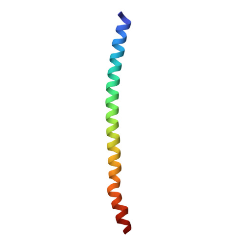
Contraction in striated and cardiac muscles is regulated by the motions of a Ca(2+)-sensitive tropomyosin/troponin switch. In contrast, troponin is absent in other muscle types and in nonmuscle cells, and actomyosin regulation is myosin-linked. Here we report an unusual crystal structure at 2.7 A of the C-terminal 31 residues of rat striated-muscle alpha-tropomyosin (preceded by a fragment of the GCN4 leucine zipper). The C-terminal 22 residues (263-284) of the structure do not form a two-stranded alpha-helical coiled coil as does the rest of the molecule, but here the alpha-helices splay apart and are stabilized by the formation of a tail-to-tail dimer with a symmetry-related molecule. The site of splaying involves a small group of destabilizing core residues that is present only in striated muscle tropomyosin isoforms. These results reveal a specific recognition site for troponin T and clarify the physical basis for the unique regulatory mechanism of striated muscles.
- Rosenstiel Basic Medical Sciences Research Center, Brandeis University, Waltham, MA 02454-9110, USA.
Organizational Affiliation: