1KP8 | pdb_00001kp8

Structural Basis for GroEL-assisted Protein Folding from the Crystal Structure of (GroEL-KMgATP)14 at 2.0 A Resolution


Experimental Data Snapshot

  • Method: X-RAY DIFFRACTION
  • Resolution: 2.00 Å
  • R-Value Free: 
    0.258 (Depositor), 0.256 (DCC) 
  • R-Value Work: 
    0.243 (Depositor), 0.241 (DCC) 

wwPDB Validation 3D Report Full Report

Validation slider image for 1KP8

Ligand Structure Quality Assessment 


This is version 1.5 of the entry. See complete history

Re-refinement Note

A newer entry is available that reflects an alternative modeling of the original data: 1SX3

Literature

Structural Basis for GroEL-assisted Protein Folding from the Crystal Structure of (GroEL-KMgATP)14 at 2.0 A Resolution

Wang, J.Boisvert, D.C.

(2003) J Mol Biology 327: 843-855

  • DOI: https://doi.org/10.1016/s0022-2836(03)00184-0
  • Primary Citation Related Structures: 
    1KP8

  • PubMed Abstract: 

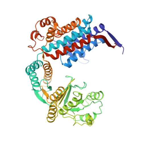
    Nucleotide regulates the affinity of the bacterial chaperonin GroEL for protein substrates. GroEL binds protein substrates with high affinity in the absence of ATP and with low affinity in its presence. We report the crystal structure of (GroEL-KMgATP)(14) refined to 2.0 A resolution in which the ATP triphosphate moiety is directly coordinated by both K(+) and Mg(2+). Upon the binding of KMgATP, we observe previously unnoticed domain rotations and a 102 degrees rotation of the apical domain surface helix I. Two major consequences are a large lateral displacement of, and a dramatic reduction of hydrophobicity in, the apical domain surface. These results provide a basis for the nucleotide-dependent regulation of protein substrate binding and suggest a mechanism for GroEL-assisted protein folding by forced unfolding.


  • Organizational Affiliation
    • Department of Molecular Biophysics and Biochemistry Yale University, 266 Whitney Avenue, Bass Center, Room 418, New Haven, CT 06520-8114, USA. wang@mail.csb.yale.edu

Macromolecule Content 

  • Total Structure Weight: 810.23 kDa 
  • Atom Count: 57,085 
  • Modeled Residue Count: 7,350 
  • Deposited Residue Count: 7,658 
  • Unique protein chains: 1

Macromolecules

Find similar proteins by:|  3D Structure
Entity ID: 1
MoleculeChains  Sequence LengthOrganismDetailsImage
groEL protein
A, B, C, D, E
A, B, C, D, E, F, G, H, I, J, K, L, M, N
547Escherichia coliMutation(s): 0 
EC: 5.6.1.7
UniProt
Find proteins for P0A6F5 (Escherichia coli (strain K12))
Explore P0A6F5 
Go to UniProtKB:  P0A6F5
Entity Groups
Sequence Clusters30% Identity50% Identity70% Identity90% Identity95% Identity100% Identity
UniProt GroupP0A6F5
Sequence Annotations
Expand
Reference Sequence

Small Molecules

Ligands 4 Unique
IDChains Name / Formula / InChI Key2D Diagram3D Interactions
AGS

Query on AGS


Download:Ideal Coordinates CCD File
AB [auth H]
BC [auth N]
DA [auth C]
DB [auth I]
HA [auth D]
AB [auth H],
BC [auth N],
DA [auth C],
DB [auth I],
HA [auth D],
IB [auth J],
NA [auth E],
NB [auth K],
RA [auth F],
RB [auth L],
T [auth A],
VA [auth G],
WB [auth M],
Y [auth B]
PHOSPHOTHIOPHOSPHORIC ACID-ADENYLATE ESTER
C10 H16 N5 O12 P3 S
NLTUCYMLOPLUHL-KQYNXXCUSA-N
SO4

Query on SO4


Download:Ideal Coordinates CCD File
AA [auth C]
EB [auth J]
FB [auth J]
JA [auth E]
JB [auth K]
AA [auth C],
EB [auth J],
FB [auth J],
JA [auth E],
JB [auth K],
KA [auth E],
KB [auth K],
O [auth A],
OA [auth F],
OB [auth L],
P [auth A],
Q [auth A],
SA [auth G],
SB [auth M],
TB [auth M],
U [auth B],
V [auth B],
WA [auth H],
XA [auth H],
XB [auth N],
YB [auth N],
Z [auth C]
SULFATE ION
O4 S
QAOWNCQODCNURD-UHFFFAOYSA-L
K

Query on K


Download:Ideal Coordinates CCD File
AC [auth N]
CA [auth C]
CB [auth I]
EA [auth D]
GA [auth D]
AC [auth N],
CA [auth C],
CB [auth I],
EA [auth D],
GA [auth D],
HB [auth J],
IA [auth E],
MA [auth E],
MB [auth K],
QA [auth F],
QB [auth L],
S [auth A],
UA [auth G],
VB [auth M],
X [auth B],
ZA [auth H]
POTASSIUM ION
K
NPYPAHLBTDXSSS-UHFFFAOYSA-N
MG

Query on MG


Download:Ideal Coordinates CCD File
BA [auth C]
BB [auth I]
FA [auth D]
GB [auth J]
LA [auth E]
BA [auth C],
BB [auth I],
FA [auth D],
GB [auth J],
LA [auth E],
LB [auth K],
PA [auth F],
PB [auth L],
R [auth A],
TA [auth G],
UB [auth M],
W [auth B],
YA [auth H],
ZB [auth N]
MAGNESIUM ION
Mg
JLVVSXFLKOJNIY-UHFFFAOYSA-N

Experimental Data & Validation

Experimental Data

  • Method: X-RAY DIFFRACTION
  • Resolution: 2.00 Å
  • R-Value Free:  0.258 (Depositor), 0.256 (DCC) 
  • R-Value Work:  0.243 (Depositor), 0.241 (DCC) 
Space Group: P 1 21 1
Unit Cell:
Length ( Å )Angle ( ˚ )
a = 135.571α = 90
b = 260.112β = 101.14
c = 150.2γ = 90
Software Package:
Software NamePurpose
CNSrefinement
DENZOdata reduction
SCALEPACKdata scaling

Structure Validation

View Full Validation Report


Ligand Structure Quality Assessment 


Entry History 

Deposition Data

  • Released Date: 2003-03-25 
  • Deposition Author(s): Wang, J.
  • This entry supersedes: 1DER

Revision History  (Full details and data files)

  • Version 1.0: 2003-03-25
    Type: Initial release
  • Version 1.1: 2008-04-27
    Changes: Version format compliance
  • Version 1.2: 2011-07-13
    Changes: Version format compliance
  • Version 1.3: 2014-03-26
    Changes: Non-polymer description, Version format compliance
  • Version 1.4: 2017-10-11
    Changes: Refinement description
  • Version 1.5: 2024-02-14
    Changes: Data collection, Database references, Derived calculations