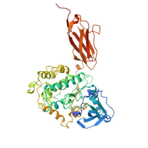
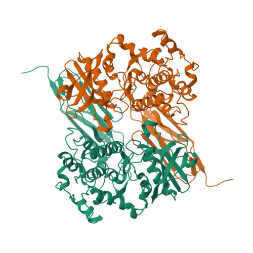
1KOA

TWITCHIN KINASE FRAGMENT (C.ELEGANS), AUTOREGULATED PROTEIN KINASE AND IMMUNOGLOBULIN DOMAINS


Experimental Data Snapshot

  • Method: X-RAY DIFFRACTION
  • Resolution: 3.30 Å
  • R-Value Free: 0.351 
  • R-Value Work: 0.251 
  • R-Value Observed: 0.251 

wwPDB Validation   3D Report Full Report


This is version 1.3 of the entry. See complete history


Literature

Giant protein kinases: domain interactions and structural basis of autoregulation.

Kobe, B.Heierhorst, J.Feil, S.C.Parker, M.W.Benian, G.M.Weiss, K.R.Kemp, B.E.

(1996) EMBO J 15: 6810-6821

  • Primary Citation of Related Structures:  
    1KOA, 1KOB

  • PubMed Abstract: 

    The myosin-associated giant protein kinases twitchin and titin are composed predominantly of fibronectin- and immunoglobulin-like modules. We report the crystal structures of two autoinhibited twitchin kinase fragments, one from Aplysia and a larger fragment from Caenorhabditis elegans containing an additional C-terminal immunoglobulin-like domain. The structure of the longer fragment shows that the immunoglobulin domain contacts the protein kinase domain on the opposite side from the catalytic cleft, laterally exposing potential myosin binding residues. Together, the structures reveal the cooperative interactions between the autoregulatory region and the residues from the catalytic domain involved in protein substrate binding, ATP binding, catalysis and the activation loop, and explain the differences between the observed autoinhibitory mechanism and the one found in the structure of calmodulin-dependent kinase I.


  • Organizational Affiliation

    St. Vincent's Institute of Medical Research, Fitzroy, Victoria, Australia.


Macromolecules
Find similar proteins by:  (by identity cutoff)  |  3D Structure
Entity ID: 1
MoleculeChains Sequence LengthOrganismDetailsImage
TWITCHIN491Caenorhabditis elegansMutation(s): 0 
EC: 2.7.11.1
UniProt
Find proteins for Q23551 (Caenorhabditis elegans)
Explore Q23551 
Go to UniProtKB:  Q23551
Entity Groups  
Sequence Clusters30% Identity50% Identity70% Identity90% Identity95% Identity100% Identity
UniProt GroupQ23551
Sequence Annotations
Expand
  • Reference Sequence
Experimental Data & Validation

Experimental Data

  • Method: X-RAY DIFFRACTION
  • Resolution: 3.30 Å
  • R-Value Free: 0.351 
  • R-Value Work: 0.251 
  • R-Value Observed: 0.251 
  • Space Group: I 21 3
Unit Cell:
Length ( Å )Angle ( ˚ )
a = 195.5α = 90
b = 195.5β = 90
c = 195.5γ = 90
Software Package:
Software NamePurpose
X-PLORmodel building
X-PLORrefinement
DENZOdata reduction
SCALEPACKdata scaling
X-PLORphasing

Structure Validation

View Full Validation Report


Entry History 

Deposition Data

Revision History  (Full details and data files)

  • Version 1.0: 1997-03-12
    Type: Initial release
  • Version 1.1: 2008-03-24
    Changes: Version format compliance
  • Version 1.2: 2011-07-13
    Changes: Version format compliance
  • Version 1.3: 2024-02-14
    Changes: Data collection, Database references, Other