1KN3 | pdb_00001kn3

Murine PEBP-2 (phosphatidylethanolamine-binding protein-2)


Experimental Data Snapshot

  • Method: X-RAY DIFFRACTION
  • Resolution: 1.80 Å
  • R-Value Free: 
    0.267 (Depositor), 0.266 (DCC) 
  • R-Value Work: 
    0.210 (Depositor), 0.215 (DCC) 
  • R-Value Observed: 
    0.213 (Depositor) 

Starting Model: experimental
View more details

wwPDB Validation 3D Report Full Report

Validation slider image for 1KN3

This is version 1.3 of the entry. See complete history

Literature

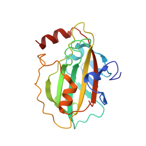
The crystal structure of PEBP-2, a homologue of the PEBP/RKIP family.

Simister, P.C.Banfield, M.J.Brady, R.L.

(2002) Acta Crystallogr D Biol Crystallogr 58: 1077-1080

  • DOI: https://doi.org/10.1107/s090744490200522x
  • Primary Citation Related Structures: 
    1KN3

  • PubMed Abstract: 

    Proteins from the PEBP (phosphatidylethanolamine-binding protein) family have been identified in a wide variety of species and are thought to regulate a range of intracellular signalling cascades. The rat homologue (known as RKIP; Raf-1 kinase inhibitor protein) has been shown to negatively regulate the MAP kinase pathway through formation of inhibitory complexes with Raf-1 and MEK. The crystal structure of a new, murine member of the PEBP family, termed mPEBP-2, has been determined. On the basis of amino-acid homology, mPEBP-2 belongs to a distinct subset of the mammalian PEBP proteins. Nonetheless, mPEBP-2 is seen to be very similar in structure to other PEBP proteins from human, bovine and plant sources. Regions of distinctive sequence associated with the PEBP-2 subset are discussed with reference to this structure.


  • Organizational Affiliation
    • Department of Biochemistry, University of Bristol, Bristol BS8 1TD, England.

Macromolecule Content 

  • Total Structure Weight: 20.78 kDa 
  • Atom Count: 1,574 
  • Modeled Residue Count: 180 
  • Deposited Residue Count: 183 
  • Unique protein chains: 1

Macromolecules

Find similar proteins by:|  3D Structure
Entity ID: 1
MoleculeChains  Sequence LengthOrganismDetailsImage
Phosphatidylethanolamine Binding Protein-2183Mus musculusMutation(s): 0 
Gene Names: pebp-2
UniProt
Find proteins for Q8VIN1 (Mus musculus)
Explore Q8VIN1 
Go to UniProtKB:  Q8VIN1
Entity Groups
Sequence Clusters30% Identity50% Identity70% Identity90% Identity95% Identity100% Identity
UniProt GroupQ8VIN1
Sequence Annotations
Expand
Reference Sequence

Experimental Data & Validation

Experimental Data

  • Method: X-RAY DIFFRACTION
  • Resolution: 1.80 Å
  • R-Value Free:  0.267 (Depositor), 0.266 (DCC) 
  • R-Value Work:  0.210 (Depositor), 0.215 (DCC) 
  • R-Value Observed: 0.213 (Depositor) 
Space Group: P 21 21 21
Unit Cell:
Length ( Å )Angle ( ˚ )
a = 40.816α = 90
b = 51.401β = 90
c = 79.765γ = 90
Software Package:
Software NamePurpose
AMoREphasing
REFMACrefinement
DENZOdata reduction
SCALEPACKdata scaling

Structure Validation

View Full Validation Report


Entry History 

Deposition Data

Revision History  (Full details and data files)

  • Version 1.0: 2002-06-12
    Type: Initial release
  • Version 1.1: 2008-04-27
    Changes: Version format compliance
  • Version 1.2: 2011-07-13
    Changes: Advisory, Version format compliance
  • Version 1.3: 2023-08-16
    Changes: Data collection, Database references, Refinement description