1KH8 | pdb_00001kh8

Structure of a cis-proline (P114) to glycine variant of Ribonuclease A


Experimental Data Snapshot

  • Method: X-RAY DIFFRACTION
  • Resolution: 2.00 Å
  • R-Value Free: 
    0.219 (Depositor), 0.222 (DCC) 
  • R-Value Work: 
    0.195 (Depositor), 0.195 (DCC) 

Starting Model: experimental
View more details

wwPDB Validation 3D Report Full Report

Validation slider image for 1KH8

This is version 1.5 of the entry. See complete history

Literature

The crystal structure of the cis-proline to glycine variant (P114G) of ribonuclease A.

Schultz, D.A.Friedman, A.M.White, M.A.Fox, R.O.

(2005) Protein Sci 14: 2862-2870

  • DOI: https://doi.org/10.1110/ps.051610505
  • Primary Citation Related Structures: 
    1KH8

  • PubMed Abstract: 

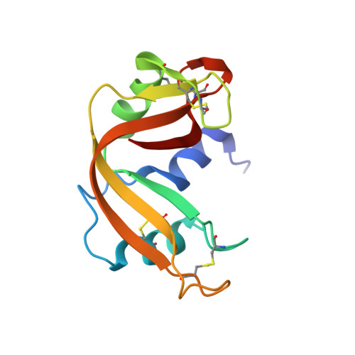
    Replacement of a cis-proline by glycine at position 114 in ribonuclease A leads to a large decrease in thermal stability and simplifies the refolding kinetics. A crystallographic approach was used to determine whether the decrease in thermal stability results from the presence of a cis glycine peptide bond, or from a localized structural rearrangement caused by the isomerization of the mutated cis 114 peptide bond. The structure was solved at 2.0 A resolution and refined to an R-factor of 19.5% and an R(free) of 21.9%. The overall conformation of the protein was similar to that of wild-type ribonuclease A; however, there was a large localized rearrangement of the mutated loop (residues 110-117-a 9.3 A shift of the Calpha atom of residue 114). The peptide bond before Gly114 is in the trans configuration. Interestingly, a large anomalous difference density was found near residue 114, and was attributed to a bound cesium ion present in the crystallization experiment. The trans isomeric configuration of the peptide bond in the folded state of this mutant is consistent with the refolding kinetics previously reported, and the associated protein conformational change provides an explanation for the decreased thermal stability.


  • Organizational Affiliation
    • Department of Physics, University of California-San Diego, La Jolla, CA 92093, USA.

Macromolecule Content 

  • Total Structure Weight: 14.03 kDa 
  • Atom Count: 1,024 
  • Modeled Residue Count: 125 
  • Deposited Residue Count: 125 
  • Unique protein chains: 1

Macromolecules

Find similar proteins by:|  3D Structure
Entity ID: 1
MoleculeChains  Sequence LengthOrganismDetailsImage
pancreatic ribonuclease A125Bos taurusMutation(s): 1 
EC: 3.1.27.5 (PDB Primary Data), 4.6.1.18 (UniProt)
UniProt
Find proteins for P61823 (Bos taurus)
Explore P61823 
Go to UniProtKB:  P61823
Entity Groups
Sequence Clusters30% Identity50% Identity70% Identity90% Identity95% Identity100% Identity
UniProt GroupP61823
Sequence Annotations
Expand
Reference Sequence

Experimental Data & Validation

Experimental Data

  • Method: X-RAY DIFFRACTION
  • Resolution: 2.00 Å
  • R-Value Free:  0.219 (Depositor), 0.222 (DCC) 
  • R-Value Work:  0.195 (Depositor), 0.195 (DCC) 
Space Group: P 43 21 2
Unit Cell:
Length ( Å )Angle ( ˚ )
a = 40.87α = 90
b = 40.87β = 90
c = 129.25γ = 90
Software Package:
Software NamePurpose
DENZOdata reduction
SCALEPACKdata scaling
X-PLORmodel building
CNSrefinement
X-PLORphasing

Structure Validation

View Full Validation Report


Entry History 

Deposition Data

Revision History  (Full details and data files)

  • Version 1.0: 2003-06-17
    Type: Initial release
  • Version 1.1: 2008-04-27
    Changes: Version format compliance
  • Version 1.2: 2011-07-13
    Changes: Version format compliance
  • Version 1.3: 2021-10-27
    Changes: Database references, Derived calculations
  • Version 1.4: 2024-04-03
    Changes: Data collection, Refinement description
  • Version 1.5: 2024-11-13
    Changes: Structure summary