1KFP | pdb_00001kfp

Solution structure of the antimicrobial 18-residue gomesin


Experimental Data Snapshot

  • Method: SOLUTION NMR
  • Conformers Calculated: 200 
  • Conformers Submitted: 20 
  • Selection Criteria: structures with acceptable covalent geometry,structures with favorable non-bond energy,structures with the least restraint violations,structures with the lowest energy 

wwPDB Validation 3D Report Full Report

Validation slider image for 1KFP

This is version 2.1 of the entry. See complete history

Literature

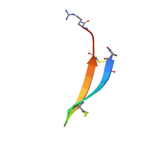
The solution structure of gomesin, an antimicrobial cysteine-rich peptide from the spider.

Mandard, N.Bulet, P.Caille, A.Daffre, S.Vovelle, F.

(2002) Eur J Biochem 269: 1190-1198

  • DOI: https://doi.org/10.1046/j.0014-2956.2002.02760.x
  • Primary Citation Related Structures: 
    1KFP

  • PubMed Abstract: 

    Gomesin is the first peptide isolated from spider exhibiting antimicrobial activities. This highly cationic peptide is composed of 18 amino-acid residues including four cysteines forming two disulfide linkages. The solution structure of gomesin has been determined using proton two-dimensional NMR (2D-NMR) and restrained molecular dynamics calculations. The global fold of gomesin consists in a well-resolved two-stranded antiparallel betasheet connected by a noncanonical betaturn. A comparison between the structures of gomesin and protegrin-1 from porcine and androctonin from scorpion outlines several common features in the distribution of hydrophobic and hydrophilic residues. The N- and C-termini, the betaturn and one face of the betasheet are hydrophilic, but the hydrophobicity of the other face depends on the peptide. The similarities suggest that the molecules interact with membranes in an analogous manner. The importance of the intramolecular disulfide bridges in the biological activity of gomesin is being investigated.


  • Organizational Affiliation
    • Centre de Biophysique Moléculaire, CNRS, Orléans, France.

Macromolecule Content 

  • Total Structure Weight: 2.28 kDa 
  • Atom Count: 156 
  • Modeled Residue Count: 19 
  • Deposited Residue Count: 19 
  • Unique protein chains: 1

Macromolecules

Find similar proteins by:  Sequence   |   3D Structure  
Entity ID: 1
MoleculeChains  Sequence LengthOrganismDetailsImage
GOMESIN19Acanthoscurria gomesianaMutation(s): 0 
Membrane Entity: Yes 
UniProt
Find proteins for P82358 (Acanthoscurria gomesiana)
Explore P82358 
Go to UniProtKB:  P82358
Entity Groups
Sequence Clusters30% Identity50% Identity70% Identity90% Identity95% Identity100% Identity
UniProt GroupP82358
Sequence Annotations
Expand
Reference Sequence

Small Molecules

Modified Residues  1 Unique
IDChains TypeFormula2D DiagramParent
PCA
Query on PCA
A
L-PEPTIDE LINKINGC5 H7 N O3GLN

Experimental Data & Validation

Experimental Data

  • Method: SOLUTION NMR
  • Conformers Calculated: 200 
  • Conformers Submitted: 20 
  • Selection Criteria: structures with acceptable covalent geometry,structures with favorable non-bond energy,structures with the least restraint violations,structures with the lowest energy 

Structure Validation

View Full Validation Report


Entry History 

Deposition Data

Revision History  (Full details and data files)

  • Version 1.0: 2002-04-10
    Type: Initial release
  • Version 1.1: 2008-04-27
    Changes: Version format compliance
  • Version 1.2: 2011-07-13
    Changes: Version format compliance
  • Version 2.0: 2019-12-25
    Changes: Database references, Derived calculations, Polymer sequence, Source and taxonomy
  • Version 2.1: 2024-10-16
    Changes: Data collection, Database references, Derived calculations, Structure summary